The Benefits of optomap Imaging
What is Ultra-widefield retinal imaging?
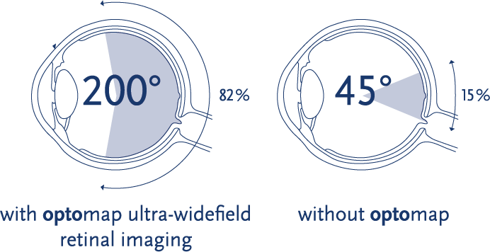
With traditional, small-field, and even widefield retinal imaging, only 10-100⁰ of the retina can be captured in a single image. optomap is the only true, clinically validated, ultra-widefield retinal image that can capture 82% or 200⁰ of the retina, in a single capture, in each imaging modality – an increase of 50% over the next closest imaging device1. With optomap auto-montage, up to 97% or 220⁰ of the retina can be imaged with the multi-capture, montaging functionality.
optomap offers unparalleled views of the retina which provide eye care professionals with the following:
- The only anatomically correct 200⁰ or 82% image of the retina
- Simultaneous view of the central pole, mid-periphery and periphery
- Integrated hardware & software platform
- Multiple retinal imaging modalities to see more, discover more, and treat more patient diseases and pathologies, more effectively
- With more than 2500 published and ongoing clinical trials, as well as thousands of case studies and testimonials, show the long-term value of optomap imaging in diagnosis, treatment planning, and patient engagement

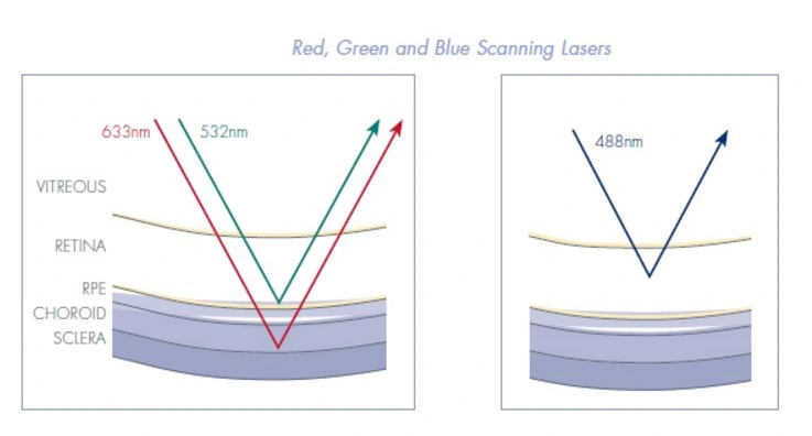
Unlike full-spectrum white light used in conventional devices, optomap technology incorporates low-powered laser wavelengths that scan simultaneously. This allows review of the retinal substructures in their individual laser separations
- Green laser (532 nm) scans from the sensory retina to the pigment epithelial layers
- Red laser (633 nm) scans from the RPE to the choroid
- Blue laser (488 nm) used in fluorescein angiography procedures and color rgb imaging
- Infrared laser (802 nm) used in indocyanine green angiography procedures

See the true difference that ultra-widefield imaging makes for detecting ocular and systemic disease versus traditional imaging methods.
*Auto-montage is available for optomap color and optomap af
1 Witmer, Parlitsis, Patel, Kiss. Weill Cornell Medical College, New York, NY, USA. Clinical Ophthalmology, Feb 20, 2013