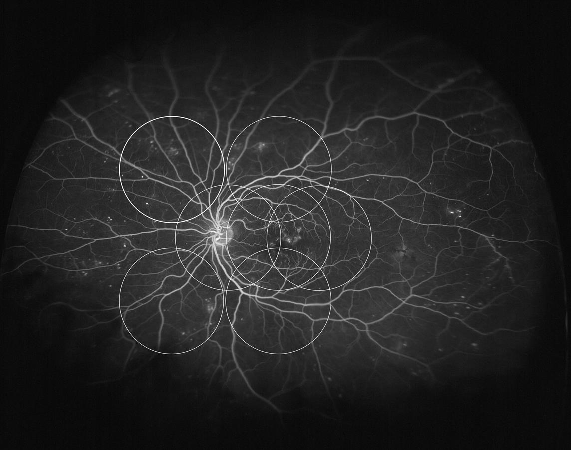
A prospective comparative study published recently in Ophthalmology found substantial agreement (80%) between Optos UWF and ETDRS 7-standard field images of eyes with DR. However, by identifying additional lesions in the retinal periphery, UWF imaging led to a more severe DR assessment in 10% of eyes, compared to assessments based on conventional photography. The authors call for further prospective study to evaluate the implications of these lesions for DR progression within different levels of severity.

optomap® fa demonstrating the peripheral changes associated with diabetic retinopathy that are not able to be visualized using ETDRS 7 standard field photography.
Silva PS, Cavallerano JD, Sun JK, Soliman AZ, Aiello LM, Aiello LP. Peripheral lesions identified by mydriatic ultrawide field imaging: Distribution and potential impact on diabetic retinopathy severity. Ophthalmology. 2013: 1-9. [Epub ahead of print]