With the peripheral retina being the location of pathology for lots of ocular diseases, such as diabetic retinopathy, chorodial masses and vasculitis, the need for advanced imaging technology is a must. As Matthew T. Witmer, MD, and Szilárd Kiss, MD, of New York City shared with Review of Ophthalmology, even though the “gold standard” for a complete retinal exam was once considered a dilated exam with indirect ophthalmoscopy and occasional scleral depression, advances in technology are making the process of evaluating and documenting the retinal periphery through photography a more practical way to conduct an exam in clinical environments.
The duo discussed four technologies that “permit imaging of the retinal periphery in a practical manner,” including Optos’ ultra-widefield (UWF) retinal imaging technology. They compared the four technologies with traditional fundus photography from The Diabetic Retinopathy Study. In that study, traditional lenses were used to capture images of the posterior pole, with the ability to fuse multiple images into a montage. The image created only covered a field of view about 75 degrees wide.
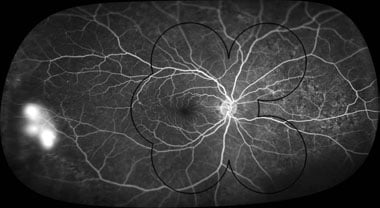
Optos’ imaging technology, on the other hand, produces images capable of a 200 degree field of view, which is equivalent to about 82.5 percent of the retina’s total surface. Not only did Optos’ UWF imaging provide a wider field of view, but it also offered a clinical benefit of producing “high-resolution fluorescein angiogram images of the retinal periphery,” which is extremely useful from a clinical standpoint for patients with retinal vascular disease. Optos’ capability of producing autofluorescence images were also viewed to be particularly helpful for disorders that have an effect on retinal pigment epithelium.

This UWF fluorescein angiogram image captured with Optos’ technology shows the right eye of a diabetic patient and demonstrates peripheral neovascularization temporally, as well as illustrating how the areas covered by standard photography would not present this finding.
Source: Review of Ophthalmology
Are you interested in learning more about the clinical benefits Optos’ UWF retinal imaging technology can provide to your practice? Contact us today to learn more about our devices and technology.