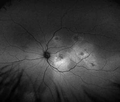
A 49 year-old Caucasian male presented a recent decrease in vision. For the past 12 years, he had a history of chronic central serous retinopathy and his vision had always recovered after each recurrence. At this visit, his visual acuity was 20/80 in the right eye and 20/40 in the left eye. The patient had no other systemic diagnosis.
Examination
optomap® af images were obtained and showed multifocal areas of hyper- and hypofluorescence reaching into the mid-periphery. The ‘gutter-like’ appearances of the broad hyperfluorescent areas are characteristic of chronic central serous retinopathy. Additionally, FD-OCT images were acquired and showed massive fibrinous exudates in the subretinal space. The patient underwent a fluorescein angiogram after optomap af images were acquired, which confirmed the diagnosis of CSR.
Discussion
Multimodal imaging was required to determine the diagnosis of CSR. The mottled appearance of the focal lesions and the broad hyperfluorescent streaks on the optomap af images are indicative of chronic central serous retinopathy as well as the “smoke-stack” leakage in the late fluorescein angiogram images. The benefit of the widefield optomap images is it can help identify patterns which are not discernible when focusing on a small retinal area only. CSR is a good example of this, particularly in Multifocal CSR. The work-up for hypercortisolism was negative and the patient was otherwise healthy, he was observed without treatment.
Conclusion
Researcher, SriniVas R. Sadda, MD, of the Doheny Eye Institute found that the optomap af images demonstrate a classic case of CSR findings. Areas of hyper- and hypofluorescence in the mid-periphery reveal the need for widefield autofluorescence imaging in CSR cases.
To learn more about how you can “See More. Treat More Effectively™,” contact us at Optos for further insight on a future partnership.
