Currently, there are more than 3 million people in the United States and over 60 million worldwide living with glaucoma, otherwise known as “the sneak thief of sight”. It is estimated that half of those with glaucoma, do not know they have it. The disease presents no symptoms and is the leading cause of irreversible blindness, taking as much as 40% of sight without notice. January has been deemed National Glaucoma Awareness Month and is an important time to spread the world about this sight-stealing disease.
What is Glaucoma?
Glaucoma is a group of eye diseases that gradually progress, stealing sight, without symptom. Glaucoma can affect people of all ages but is most prevalent in middle-aged adults and the elderly. While there is no cure, surgery or medication can slow its affects and help to prevent further vision loss. The word ‘glaucoma’ is actually an umbrella term for a group of eye diseases that damage the delicate fibers that run from your eye to your optic nerve, which is the nerve that carries information about the images your eye sees to your brain. This damage is often the result of high fluid pressure inside the eye.
What can you do?
It is important to know your risks, those at higher risk include people of African, Asian, and Hispanic descent. Other high-risk groups include: people over 60, family members of those already diagnosed, diabetics, and people who are severely nearsighted. Annual comprehensive eye exams are important to detect, prevent and treat the effects of the disease.
optomap’s role in the management of glaucoma
Results from recently published clinical studies suggest that optomap ultra-widefield (UWF™) retinal imaging may play an essential role in glaucoma management. optomap enables eyecare professionals to discover, diagnose, document and treat ocular pathology that may first present in the periphery. optomap is a high resolution single capture image of 82% or 200 degrees of the retina. Currently, the gold standard tool for glaucoma detection is a clinical examination with a dilated slit-lamp bio-microscopy carried out by a glaucoma specialist to assess the optic disc. Recent studies suggest that UWF imaging may be suitable for diagnosing glaucoma in situations where slit-lamp bio-microscopy or digital color stereoscopy are not available. Another study also confirms that optomap has almost perfect agreement with color digital stereoscopy when assessed
by a glaucoma specialist. Continued reading on these studies and additional findings here…

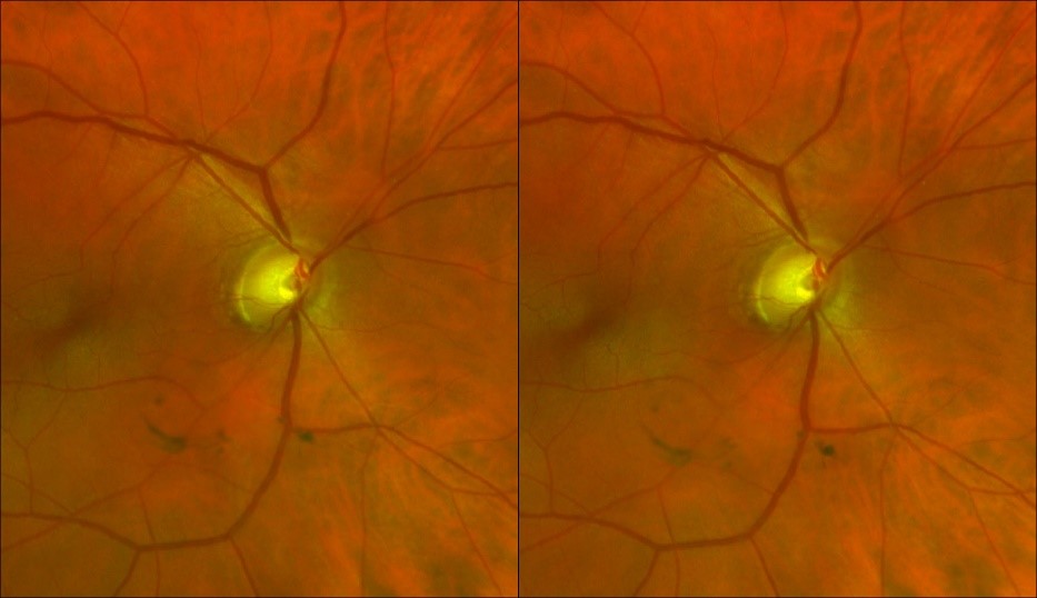
stereo pair of optic nerve head images with can be viewed using a stereo viewer, when there is suspicion of glaucoma
optomap is continuing to become a key player in the role of eye care professionals. optomap provides details needed for specialty exams, while simultaneously delivering an integrated view to the eye, as said by Dr. Savak Tymoorian, MD of Harvard Eye Associates. When Dr. Tymoorian first began using Optos technology, he employed it primary for patients presenting with flashers or floaters. While reviewing the images, he was able to pick up on more peripheral issues and early indicators of pathology. “The more I use the device, the more I appreciate this dynamic technology, I now image all my patients this way”, states Tymoorian. As a glaucoma specialist, Dr Tymoorian finds that optomap helps reassure him that he is not missing peripheral issues that could be relevant to the disease.
Recognizing January as National Glaucoma Awareness Month, allows us to shed light on glaucoma and stress the importance of protecting your sight and preventing the onset of the disease. The best way to protect your sight from glaucoma is to get a comprehensive eye examination. This way, if you have glaucoma, treatment can begin immediately.
optomap UWF imaging captures more than 80% of the retina in a single image, whereas traditional imaging methods can sometimes only reveal 10 – 15%. optomap is a fast and easy addition to a standard comprehensive eye exam. Don’t hesitate, and ask your eye care professional about optomap today.