OD Embraces UWF Technology as Important Screening Tool for Patients and Education
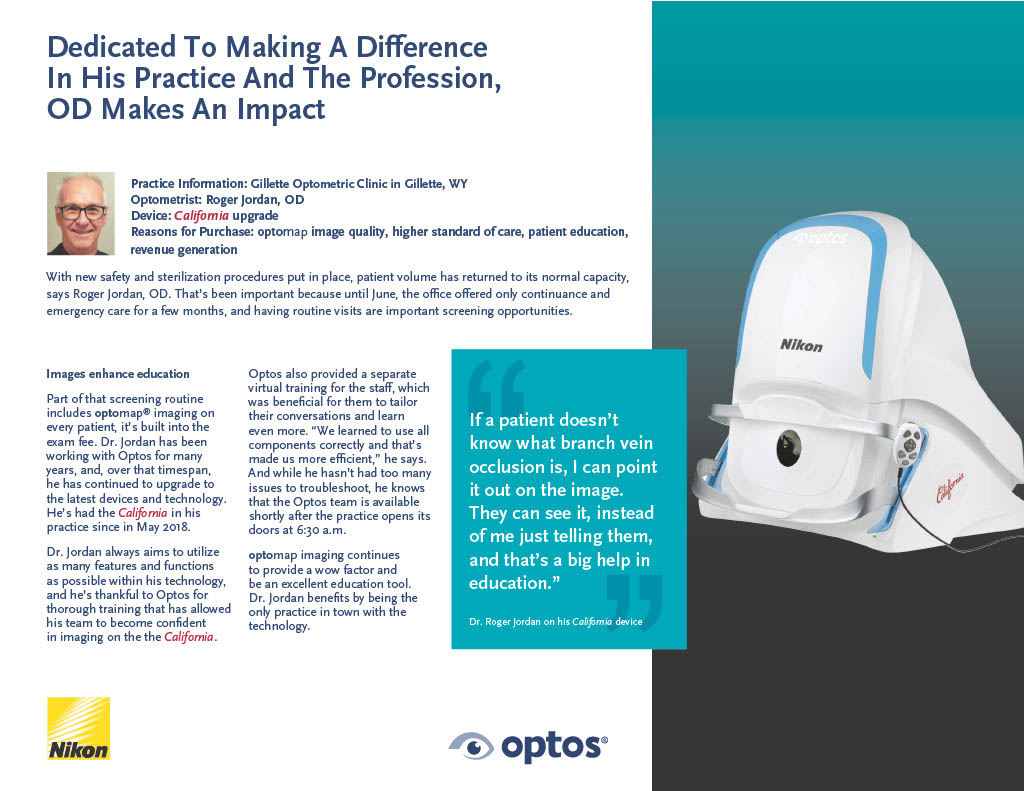
Dr. Roger Jordan of Gillette Optometry, in Gillette, WY, always aims to utilize as many features and functions as possible with technology. Dr. Jordan has been working with Optos for many years, and, over that time span, he has continued to upgrade to the latest devices and technology. He’s had California in his practice since May 2018. Dr. Jordan is most thankful to Optos for the training his team received so that they can get the most out of their latest optomap technology. Additionally, the thorough training allowed his team to become confident in imaging on their California device.
Dr. Jordan includes an optomap retinal image as a part of routine screenings during exam visits. Should a patient not understand what they are looking at, Dr. Jordan is able to easily point and explain the image. He can show a patient with diabetes, for example how the bleeding is directly impacted by their glucose levels, they are able to see for themselves rather than just telling them. This is true for new patients to the practice and more so for returning patients who are able to see a comparison year over year of their images. The impressive technology of optomap earns patient loyalty, as the practice has seen over the years.

A particular favorite feature of Dr. Jordan’s is the autofluorescence (AF) capability. optomap af imaging allows him to see changes to the RPE, often found with age-related macular degeneration (AMD), a common eye condition that causes damage to the macular and is a leading cause of vision loss among people aged 50 and older. “I see a lot more macular degeneration and pathology because I see a larger number of older patients than our younger doctors do,” he explains.
February is deemed AMD Awareness Month in order to increase awareness and education surrounding the disease. While AMD is a condition that can lead to blindness and does not yet have a cure, there are steps that patients can take to reduce their risk of progression and prevent total vision loss.
Earlier detection and treatment of AMD can prompt steps to be taken to help reduce vision loss and slow the advance of the disease. Data suggests that the retinal periphery can exhibit some important morphological changes, such as peripheral drusen and reticular pigmentary changes, which are frequently connected with the wet form of AMD. Typically, disease progression has been documented using fundus cameras that image only about 45-50% percent of the retina. By using UWF for AMD evaluation, over 80% of the retina is now analyzed to record peripheral fluorescein angiographic changes in AMD patients. Read additional information here regarding these studies or visit our website to learn more about optomap and how it helps eyecare professionals to manage eye disease.