Understanding Myopia and the Role of optomap Ultra-widefield Imaging in Early Detection and Management
Myopia, or nearsightedness, is a common refractive error in which distant objects appear blurry while near objects remain clear. This occurs when the axial length of the eye is longer or the cornea has more curvature than average eyes, causing light rays to focus in front of the retina rather than directly on it¹ ².
How Does Myopia Develop?
Myopia most often develops in childhood, particularly between the ages of 6 and 14, and can progress during adolescence. It is influenced by both genetic predisposition and environmental factors³. Children with one or both myopic parents are at significantly increased risk⁴.
Environmental contributors include:
- Prolonged near work such as reading, tablet use, and screen time
- Limited outdoor exposure, which has been shown to be protective⁵ ⁶
The structural cause—axial elongation of the eyeball—can lead to stretching and thinning of the retina and surrounding tissues, which raises the risk for serious ocular complications, especially in high myopia (defined as -6.00 D or greater or axial length >26 mm)⁷.
Potential Complications of High Myopia
- Retinal detachment⁸
- Myopic macular degeneration (MMD)⁹
- Primary open-angle glaucoma¹⁰
- Posterior subcapsular cataract¹¹
Who is Affected by Myopia?
Myopia affects individuals of all ages but is most commonly diagnosed in school-age children. In many cases, it stabilizes in the early twenties, but progression can continue into adulthood, especially in high myopia cases¹².
Global prevalence is rising rapidly. The World Health Organization and Brien Holden Vision Institute estimate that by 2050, 50% of the global population will be myopic, and 10% will have high myopia³.
In the United States, the prevalence of myopia increased from 25% in the 1970s to over 41.6% by the early 2000s¹³.
The Role of Ultra-widefield Imaging in Myopia Management
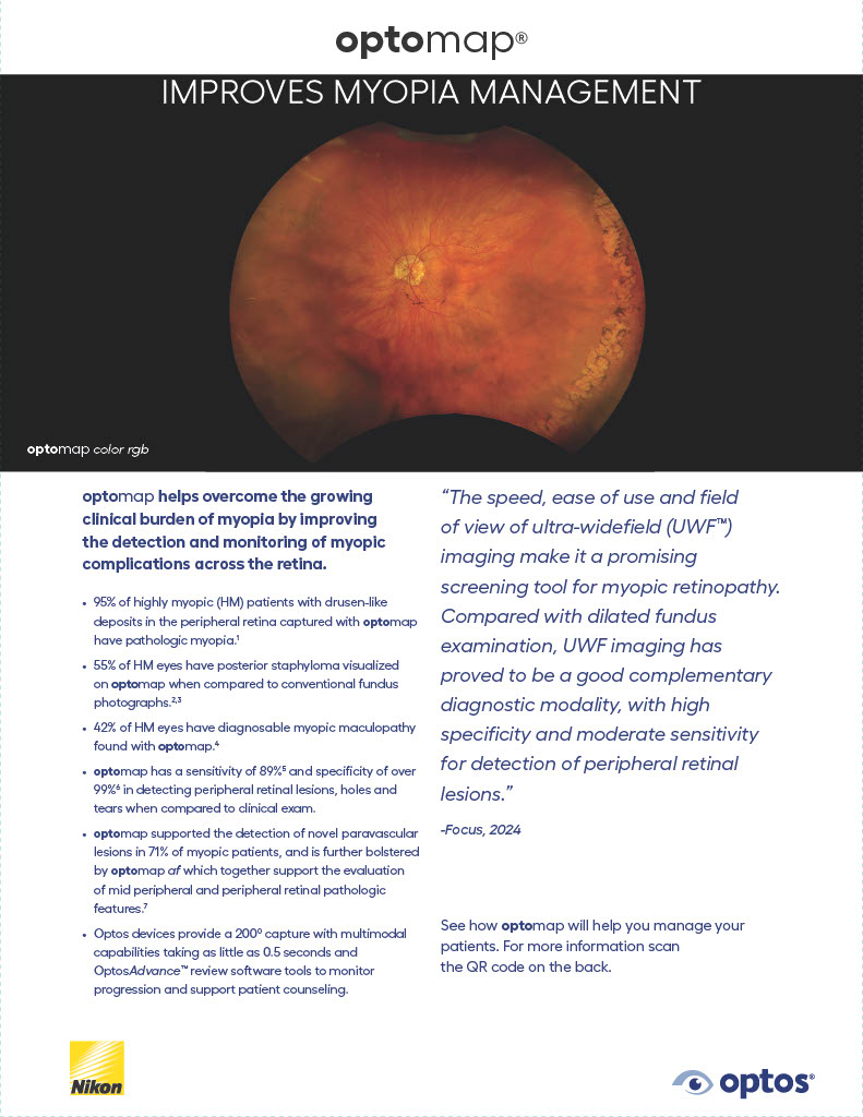
Optos ultra-widefield (UWF) retinal imaging may assist in the screening and management of myopia patients. Traditional retinal imaging systems typically capture only 30° to 50° of the retina in a single image. In contrast, optomap UWF technology can capture up to 200°, or over 80% of the retina, in one shot—without dilation. This expansive view is particularly valuable in myopia management, where peripheral retinal degenerations often present outside the central field of view.
Clinical studies have shown that optomap UWF imaging identifies peripheral retinal pathology in myopic patients that would otherwise be missed in standard dilated exams. Detecting lattice degeneration, retinal holes, or peripheral tractional changes allows clinicians to implement preventative care strategies, reducing the risk of retinal detachment and vision loss.
The MonacoPro Advantage
The MonacoPro device from Optos further elevates this approach by combining ultra-widefield imaging with spectral domain optical coherence tomography (SD-OCT) in a single device. OCT provides cross-sectional, high-resolution imaging of retinal layers, allowing for detailed visualization of structural changes associated with myopia, such as macular thinning, staphylomas, or myopic maculopathy¹⁴.
This multimodal imaging capability—UWF + OCT—enhances diagnostic precision and enables clinicians to track disease progression over time with a non-invasive, patient-friendly workflow. The MonacoPro, with its integrated system, simplifies the clinical process and improves efficiency, especially in high-volume practices and pediatric populations where cooperation may be limited¹⁴.
In the era of personalized eye care, the ability to capture comprehensive data from the macula to the periphery in one session is valuable. For patients with progressive or high myopia, this technology supports proactive decision-making, whether it’s initiating myopia control interventions (such as orthokeratology, atropine drops, or multifocal contact lenses) or scheduling more frequent monitoring based on individual risk.
As global concern over the “myopia epidemic” intensifies, Optos imaging platforms like the MonacoPro are positioned at the forefront of modern eye care. By delivering fast, detailed, and panoramic retinal assessments, these tools empower clinicians to protect vision, slow disease progression and better evaluate treatment options¹⁵.
Advancing Myopia Care
As myopia becomes a growing public health concern, early detection and personalized care are essential. optomap ultra-widefield imaging helps clinicians identify peripheral retinal changes—like lattice degeneration—that may be missed with traditional methods.
For clinical insights, case studies, and imaging strategies that support proactive management, see how Optos technology is helping eye care professionals address the challenges of progressive myopia.
To learn more about detecting retinal pathology with optomap, visit Recognizing Pathology.
References
- Flitcroft, D. I. (2012). The complex interactions of retinal, optical and environmental factors in myopia aetiology. Progress in Retinal and Eye Research, 31(6), 622–660.
- Morgan, I. G., et al. (2012). Myopia. The Lancet, 379(9827), 1739–1748.
- Holden, B. A., et al. (2016). Global prevalence of myopia and high myopia and temporal trends from 2000 through 2050. Ophthalmology, 123(5), 1036–1042.
- Tideman, J. W., et al. (2016). Association of axial length with risk of uncorrectable visual impairment for Europeans with myopia. JAMA Ophthalmology, 134(12), 1355–1363.
- Rose, K. A., et al. (2008). Outdoor activity reduces the prevalence of myopia in children. Ophthalmology, 115(8), 1279–1285.
- Wu, P. C., et al. (2013). Outdoor activity during class recess reduces myopia onset and progression in school children. Ophthalmology, 120(5), 1080–1085.
- Ohno-Matsui, K., et al. (2016). Myopia as a growing public health problem in high-prevalence countries. Retina, 36(10), 1806–1811.
- Curtin, B. J., & Karlin, D. B. (1970). Axial length measurements and fundus changes of the myopic eye. American Journal of Ophthalmology, 69(4), 695–700.
- Ohno-Matsui, K., et al. (2015). Myopic macular degeneration. Retina.
- Marcus, M. W., et al. (2001). Myopia as a risk factor for open-angle glaucoma: a systematic review and meta-analysis. Ophthalmology, 108(10), 1913–1921.
- Saw, S. M., et al. (2005). Risk factors for the progression of myopia in young Singaporean children: a cohort study. British Journal of Ophthalmology, 89(12), 1495–1500.
- Donovan, L., et al. (2012). Myopia progression rates in urban children wearing single-vision spectacles. Optometry and Vision Science, 89(1), 27–32.
- Vitale, S., et al. (2009). Increased prevalence of myopia in the United States between 1971–1972 and 1999–2004. Archives of Ophthalmology, 127(12), 1632–1639.
- Aiello. Integrating Macular Optical Coherence Tomography with Ultrawide Field Imaging in a Diabetic Retinopathy Telemedicine Program Using a Single Device. Retina. 2023.
- Ludwig CA, Moon J, Garg I, Miller JB. (2021). Ultra-Widefield Imaging for Evaluation of the Myopic Eye. Semin Ophthalmol, 36(4):185-190. doi: 10.1080/08820538.2021.1887904.