June is Cataract Awareness Month: Enhancing Retinal Care with Ultra-Widefield Imaging
In recognition of Cataract Awareness Month, Optos proudly joins Prevent Blindness America in raising awareness about cataracts—the leading cause of vision loss globally—and highlighting the critical role of ultra-widefield (UWF™) retinal imaging in modern cataract care.
Cataracts: A Growing Public Health Concern
Cataracts affect over 25 million Americans today, and this number is expected to surge to 38.5 million by 2032 as the population ages1. Cataracts cloud the eye’s natural lens, leading to symptoms such as:
- Blurry or double vision
- Increased sensitivity to light
- Difficulty seeing at night
- Faded or yellowed colors
While aging is the most common cause, other risk factors include UV exposure, diabetes, eye injuries, long-term steroid use, and smoking.
Types and Causes of Cataracts
Cataracts are classified by location (nuclear, cortical, posterior subcapsular) and cause (age-related, congenital, secondary, traumatic). They often develop in both eyes at different rates and may progress slowly, making early detection through comprehensive eye exams essential.
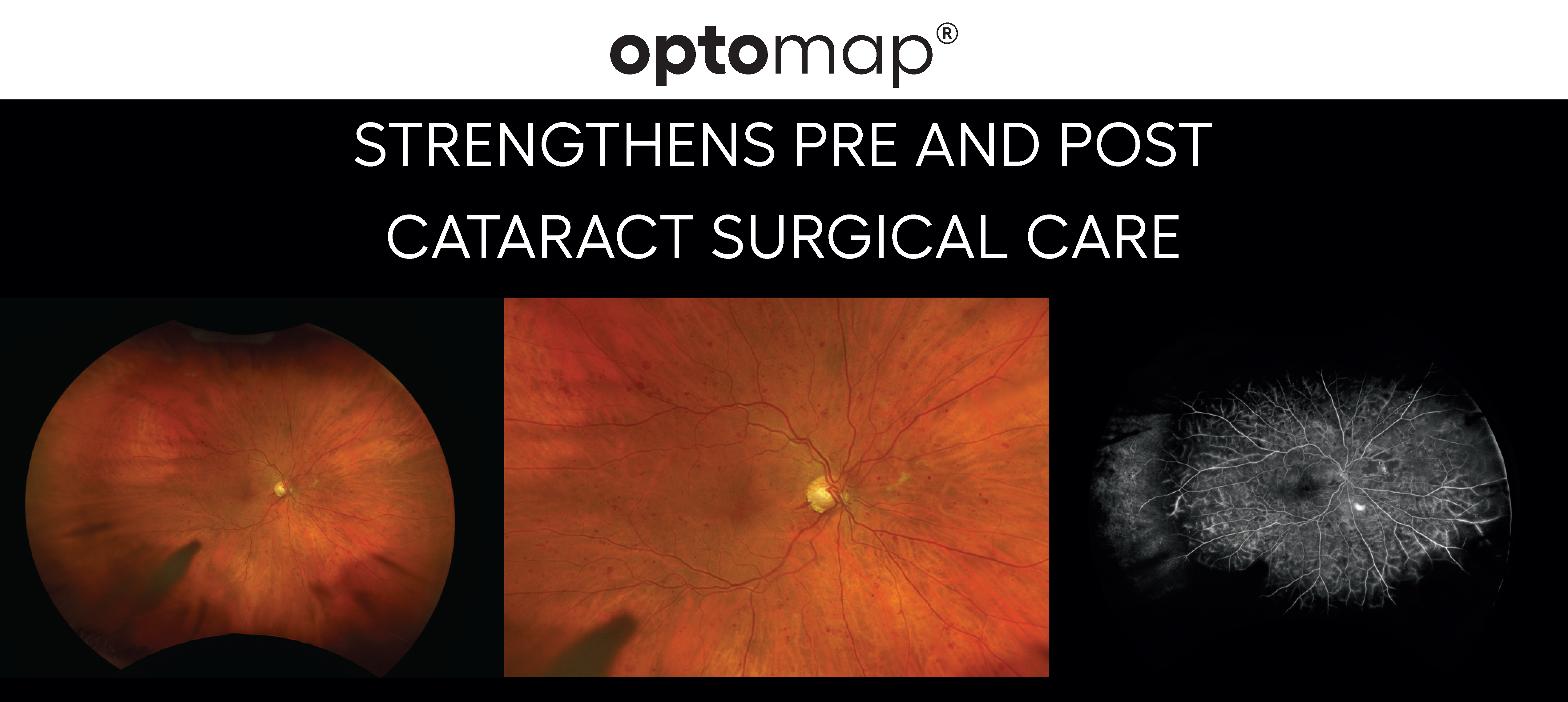
The Role of optomap® in Cataract Management
Optos’ ultra-widefield imaging technology, optomap, is increasingly integrated into cataract surgery workflows for both preoperative assessment and postoperative monitoring. This non-invasive imaging captures over 80% of the retina in a single scan—often through hazy media where traditional white light imaging struggles.
Recent clinical studies show that optomap can image through cataracts in 85% of cases, offering a clear view of the retina even when the lens is clouded.2 This capability is vital for identifying retinal pathologies such as diabetic retinopathy or macular degeneration that could impact surgical outcomes.
Clinical Evidence Supporting UWF Imaging
A 2024 paper from Assil Eye Institute and Batra Vision emphasizes the value of UWF imaging as a standard tool in cataract evaluation. The ability to detect retinal abnormalities before surgery helps avoid complications and supports better visual outcomes—especially important as younger patients increasingly opt for cataract procedures with high expectations for results.3
Key benefits of integrating optomap into cataract practices include:
- Improved diagnostic accuracy
- Streamlined clinic workflow
- Standardized documentation across practices
- Faster patient throughput
- Enhanced patient education and engagement
Efficiency and Economic Value
With optomap imaging, technicians can quickly capture high-resolution retinal images, which are instantly available for review. This reduces the need for dilated exams, shortens visit times, and allows clinicians to manage a higher volume of patients without compromising care quality.
Download the Clinical Reference Guide
For a deeper dive into the clinical applications of ultra-widefield imaging in cataract care, download the Optos Cataract Clinical Reference Guide. This comprehensive resource outlines best practices, case studies, and the latest data supporting the integration of optomap into cataract workflows. Looking for more information? Contact us to get in touch with a local Representative today.
1https://preventblindness.org/millions-of-americans-have-cataract/
2 Wendy S. ChenThomas R. FribergAndrew W. EllerCarlos Medina; Advances in Retinal Imaging of Eyes with Hazy Media: Further Studies. Invest. Ophthalmol. Vis. Sci. 2011;52(14):4036.
3 Kerry K Assil1 and V Nicholas Batra2 1. Assil Eye Institute, Beverly Hills, California, US; 2. Batra Vision, San Leandro, California, US