See More in a Single View: How Auto-Montage Enhances Imaging in OptosAdvance
See More in a Single View: How Auto-Montage Enhances Imaging in OptosAdvance
When it comes to retinal imaging, efficiency and clarity are key. That’s why OptosAdvance includes a powerful feature designed to streamline your workflow while expanding your clinical perspective: Auto-Montage.
Auto-Montage allows clinicians to automatically merge fovea and optic nerve head (ONH) images into one seamless, optomap image. The result? A single, high-quality composite image that offers a more complete retinal view without the need to toggle between multiple captures.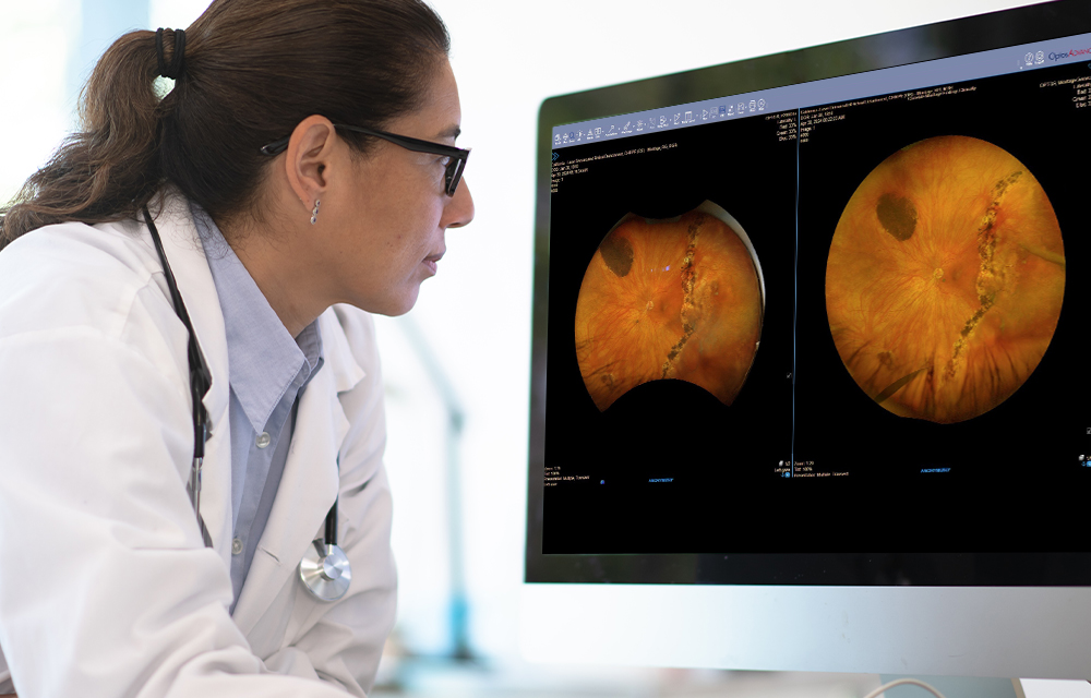
Designed for Flexibility
Available for images captured in color or green autofluorescence (af) modes, Auto-Montage offers both automatic and manual options:
Automatic Processing: The system intelligently aligns and stitches images based on preset parameters—ideal for fast-paced clinical environments.
User-Specified Selection: Clinicians can manually define the fovea and ONH images used in the montage, providing flexibility when more control is needed.
Both workflows are designed to save time while preserving image integrity. Whether you're focused on peripheral pathology, diabetic eye disease, or simply want to increase the diagnostic value of each visit, Auto-Montage helps you get more from every image.
Clinical and Operational Benefits
Auto-Montage not only enhances visualization but also contributes to a more streamlined workflow. By eliminating the need to view and compare multiple individual images, it improves clinical efficiency and supports better decision-making.
This feature is particularly valuable when using multimodal imaging strategies, allowing optomap images to be viewed in their most clinically relevant context—especially when combined with SD-OCT in or MonacoPro systems.