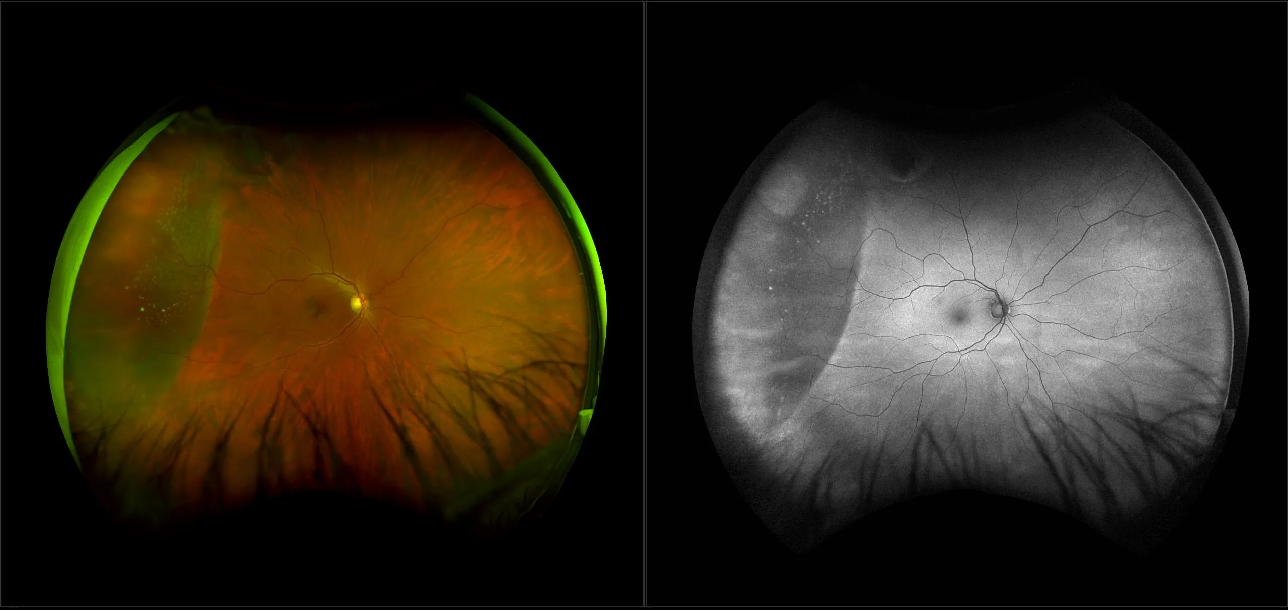
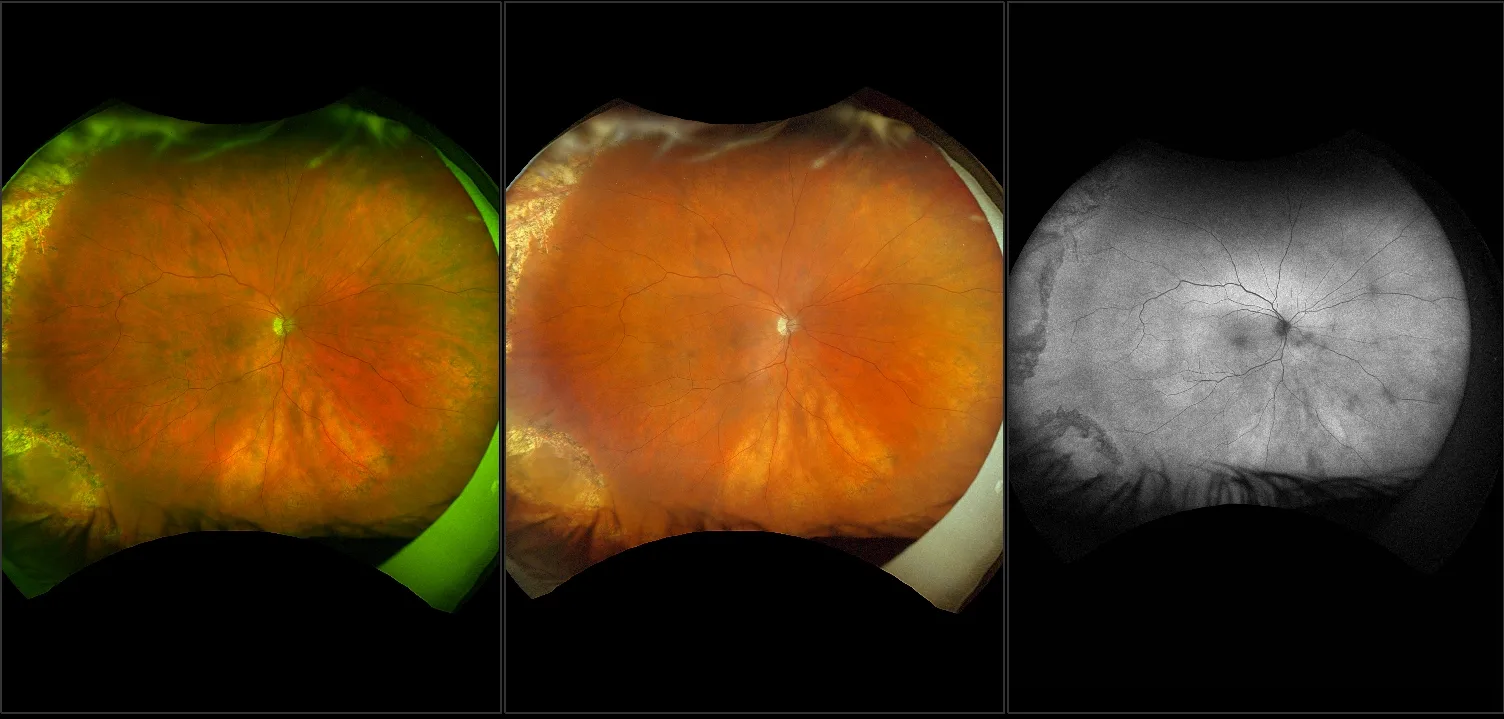
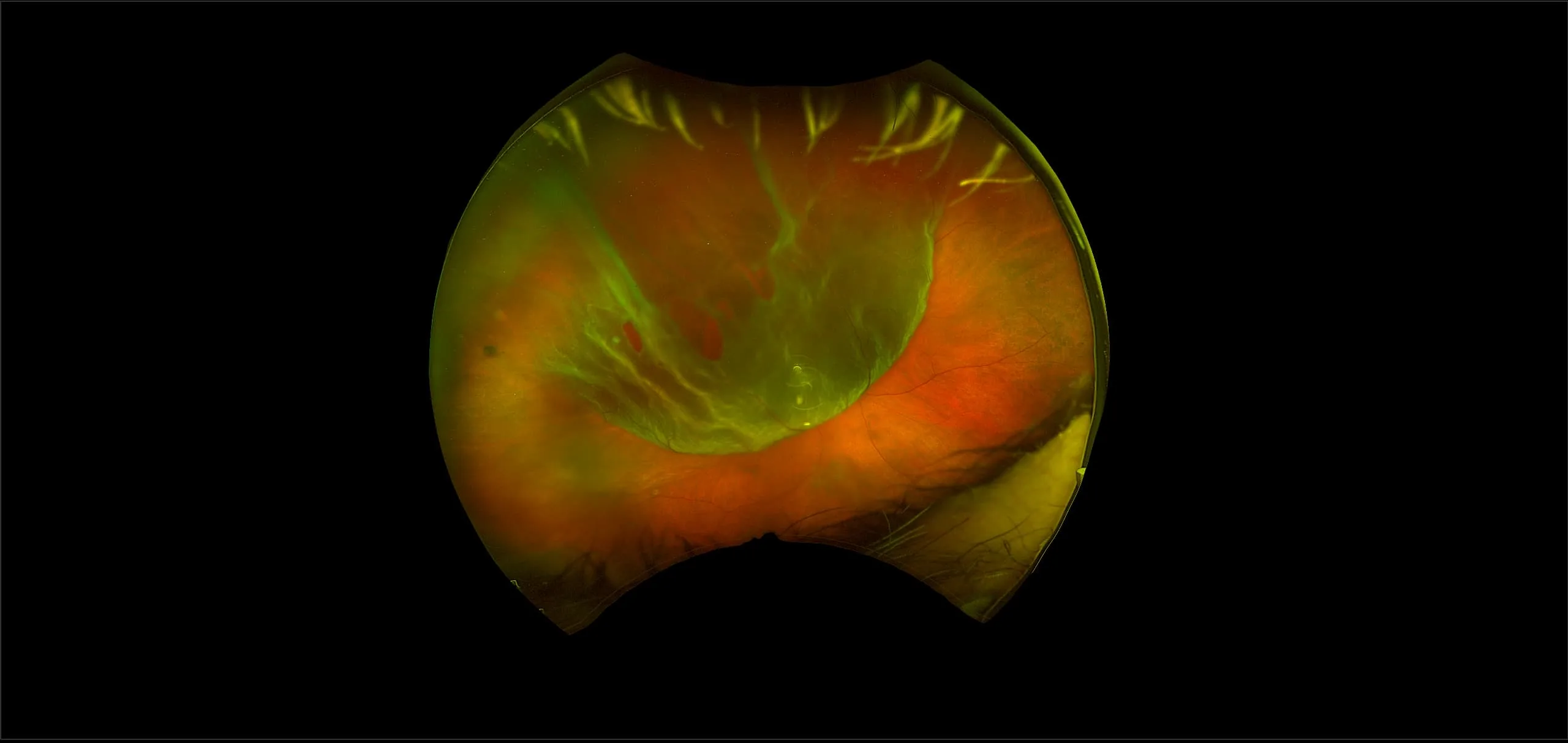
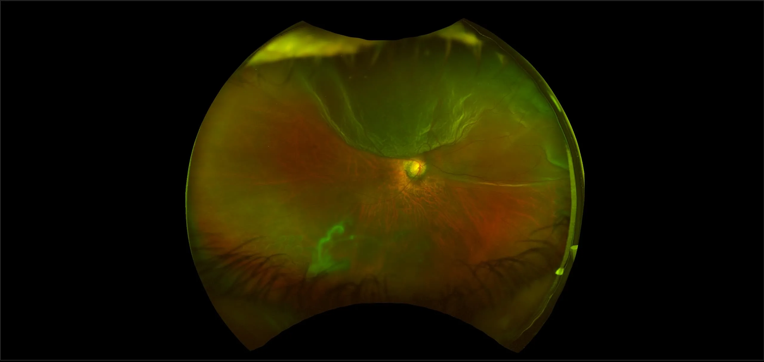
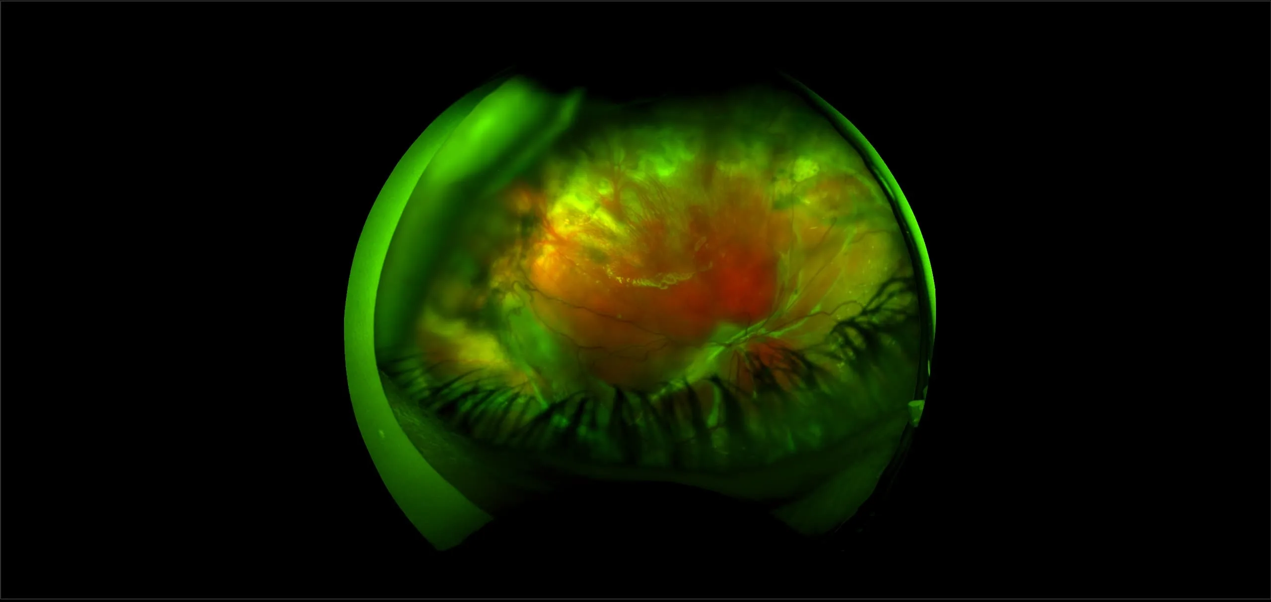
California - Retinal Detachment with Horseshoe Tear, RG, AF
What: Horseshoe tears are small areas on the retina that are torn.
Why: optomap imaging has been able to capture these tears in high resolution in both the far periphery or within the area of the vortex veins. These could have been missed with conventional limited-field imaging or during BIO (binocular indirect ophthalmoscopy) exam. Immediate treatment is required, or these tears can lead to a retinal detachment and potentially vision loss.
In the picture: optomap color image shows horseshoe tear in temporal and inferior temporal retina captured in one single image. This image may help guide treatment and can be used for documentation purposes as well as comparison for post-treatment imaging to ensure the tear has been lasered appropriately.
