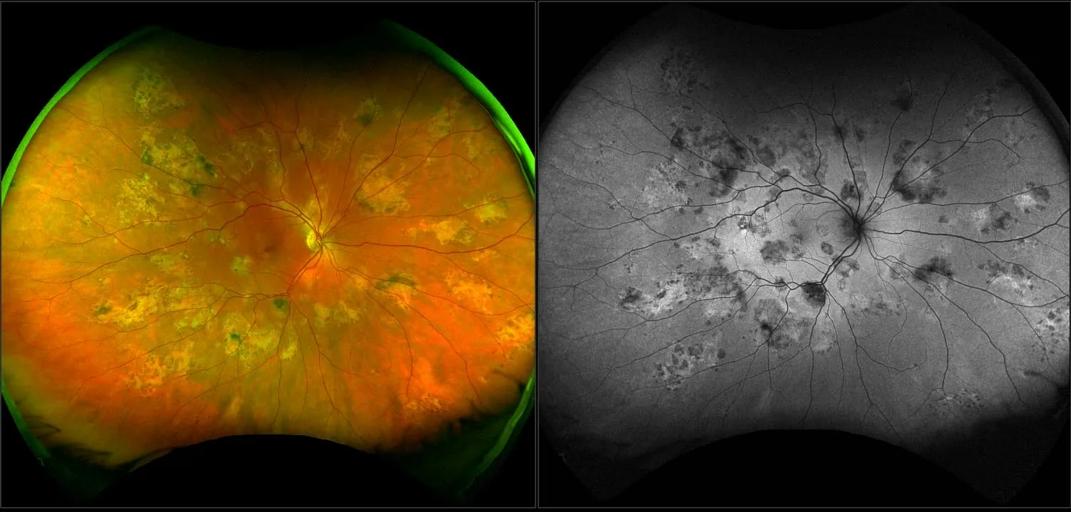
48% of Uveitis Patient's Treatment Changed with optomap

The decision to alter management was made 48% with ultra-widefield imaging compared with examination and simulated conventional FA alone.

8/16/2018

Defining Ultra-widefield - The use of consistent nomenclature when describing the FOV captured by retinal images. 

CSS - Defining UWF

“A single capture image which provides a view of the vortex veins in all four quadrants and beyond, thus meeting the widefield & ultra-widefield definitions, would offer enhanced efficiency in a real-world clinical setting versus a montage image, whether it be manual or automated.”
— Netan Choudhry M.D. FRCS(C) DABO

Choudhry, Duker, Freund, Kiss, Querques, Rosen, Sarraf, Souied, Stanga, Staurenghi, Sadda

8/1/2019

optomap Used to Guide OCT to the Far Periphery

A study published in Ophthalmology describes how ultra-widefield (UWF) optomap can be used to guide SD-OCT to image retinal features in the mid-far periphery.

Choudhry

3/16/2016

A Review of Clinical Applications and Future Trends

Optos ultra-widefield imaging has become an essential tool for the identification of peripheral retinal and vascular pathology. The high resolution and multimodal capabilities of this device are also providing new insights into a variety of disorders, even those that primarily involve the posterior pole. 

8/16/2018
Read Story

Patient Story - Brian

When 60 year-old Brian came to Belill Eye Care, in Clio, Michigan, for a non-symptomatic, routine eye exam, he never thought that his optomap would reveal that he needed to be sent for potentially sight-saving laser surgery for a tear far out in his periphery. 

Ready Story

Patient Story - David Nelson

David Nelson, OD, is a customer and like many eyecare professionals with this technology he utilizes optomap to monitor changes in his retina. With a previous retinal detachment that he had been monitoring, he went into the office early knowing that something had changed, and it had, and by 2 pm he was in sight-saving surgery.

Read Story

Patient Story - David Richardson

During a demonstration of optomap in his clinic, Dr. Richardson had an optomap taken which revealed potential cardiovascular issues. He was just about ready to go on a trip and instead went to his cardiologist. “I told him that had I not seen the image, I would have gone to my meeting in Florida and I am certain I  would have died on the beach thinking that I was having esophageal reflux or something. That optomap image literally saved my life!”

Read Story

Patient Story - Gwen

When Gwen Jacobson, 64, crossed the finish line at the 2022 Grandma’s Marathon with a sizzling 3:24 time she was ecstatic with her performance and so much more. During a routine eye exam two days before, her doctorused his state-of-the-art optomap device to take an ultra-widefield (UWF) picture of her retina. The image showed retinal tears and with her training, her doctor sent her to the Mayo clinic for laser treatment.

Read Story

Patient Story - Josh

Young Josh was asymptomatic and had no previous ocular issues and did not seem like a candidate for serious issues. Yet, the optomap image revealed extensive hemorrhages and unusual looking vessels. His optometrist suspected Coats' Disease and immediately referred Josh to a retinal specialist who confirmed that diagnosis.


Read Story

Patient Story - Luke

24 year-old Luke had a new job that came with eyecare benefits, so on a whim, he stopped in at his local LensCrafters in Chicago Ridge, IL to get fit for, and purchase contact lenses. Luke was educated on the importance of optomap in a routine eye exam. He had no symptoms but had several holes and a detachment. He was immediately sent for treatment and was relieved that he had gotten an optomap.

Read Story

Patient Story - Lynette

42 year old Lynette came to Triange Vision for a routine eye exam. Her optomap image showed bleeding in her retina which may have identified a cardiovascular issue. When she went to a local hospital screening the staff discovered that her carotid artery was 95 percent occluded on the right side and 80 percent occluded on the left side. She was immediately admitted and as she was being prepped for surgery she recalls that the surgeon remarked, “I don’t know who found this, but whoever it was, saved your life.”  Lynette credits Dr. Clark and the optomap image for alerting her to the lifethreatening issue.

Read Story

Patient Story - Mary

Mary visited her optometrist at Karns Vision Center in Knoxville, TN complaining of vision loss. Mary also let her doctor know that she was currently battling lung cancer in the event that her diagnosis and treatment were affecting her vision. Her optomap revealed she had significant pathology that was affecting her vision. Mary was advised to seek additional medical care to determine her treatment of care.

Read Story

Patient Story - Paul

Paul came to Tooele Vision Center for a routine eye exam, he knew he had a history of Coats' disease but was asymptomatic and therefore, unconcerned. His optomap image showed otherwise and he was sent to a retinal specialist for treatment. 

Tell us your story

Do you have an Optos story to tell? We want to hear how optomap may have helped save your sight or save your life, as it has with these other people, just like you. Click below to start sharing your story today.


Share Now

How can we help?

I want to receive email updates about Optos products and services