In addition to developing instruments and devices that equip optometrists and ophthalmologists with better diagnostic capabilities, we also strive to help educate practitioners by offering clinical trainings designed to provide information on the various conditions an optomap exam can detect.
Jerome Sherman, OD, FAAO and Distinguished Teaching Professor at The State University of New York, recently discussed widefield autofluorescence imaging (af) in a webinar for Optos. He describes af as a novel, noninvasive imaging procedure that has opened “a new world of diagnosis.” Throughout the webinar, Dr. Sherman discusses some of the basics of af, as well as what types of conditions can be revealed through af that can’t be detected with standard fundus photography and standard ophthalmoscopy. The webinar also includes photographic examples and some scenarios, such as the following:
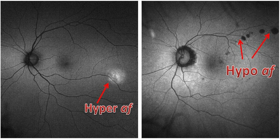
Hyperautofluorescence and Hypoautofluorescence
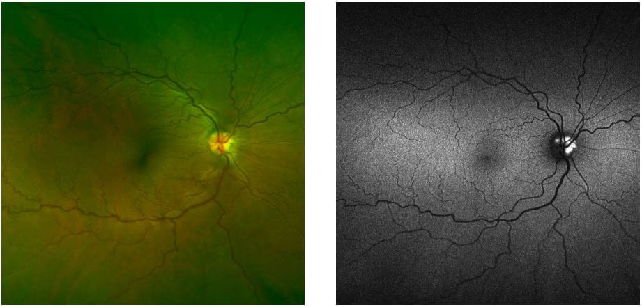
Disc Drusen revealed with af
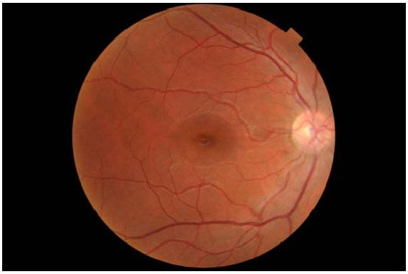
Retinal Toxicity invisible to ophthalmoscopy
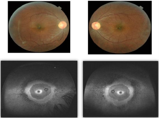
Early Retinitis Pigmentosa with Bull’s Eye Maculopathy (invisible to Ophthalmoscopy & Fundus Photography)
Please visit our website to view the entire webinar with Dr. Sherman. There, you can also find a wealth of helpful information, including clinical data and case studies on af. There you’ll also find information about retinal diagnostic instruments, medical surgical devices, and retinal imaging products like Daytona, which Dr. Sherman calls a “game changer” that is revolutionizing eyecare around the world. This compilation of information is great for practitioners already using optomap exams in their practice, as well as those who are interested in learning more about our innovative ultra-widefield retinal imaging technology.
Are you currently using widefield af in your practice? If so, how has it improved your diagnostic capabilities? Please share with us in a comment below.


