Monaco RGB
Monaco RGB takes the proven power of true color rgb ultra-widefield retinal imaging and SD-OCT together with a workflow designed for today's eye care environment. Monaco RGB combines high-quality SD-OCT imaging with up to seven imaging modalities for both eyes in just 90 seconds, simplifying the imaging experience and moving patients through the exam more efficiently. The result is a more streamlined workflow, powerful insights, and technology designed to fit the way you work.
Monaco RGB. Built for your workflow.
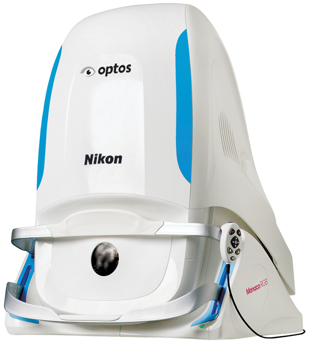

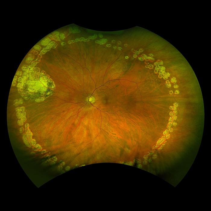
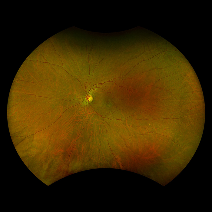
Monaco RGB is the only ultra-widefield device with integrated Spectral Domain OCT and true colour rgb retinal imaging. Monaco RGB produces a 200° single capture optomap image in less than ½ second and also provides cross-sectional 40° OCT views of retinal structures. Engineered for efficiency, Monaco RGB captures up to seven imaging modalities for both eyes in just 90 seconds, optimising workflow, streamlining diagnostics, and delivering superior patient care.
Monaco RGB offers the following benefits:
Image Capture Features
- Combined 200° single capture optomap imaging with Spectral Domain OCT
(SD-OCT) has been reported to support the detection of 29.4% more macular
pathology compared to fundus imaging alone1 - Non-Mydriatic, with minimum 2mm pupil1
- cSLO imaging through most cataracts2
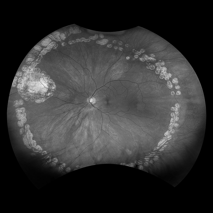
- 4-in-1 Colour DepthTM Imaging provides a true colour image with clinical data from discrete channels from the retinal surface, through the choroid
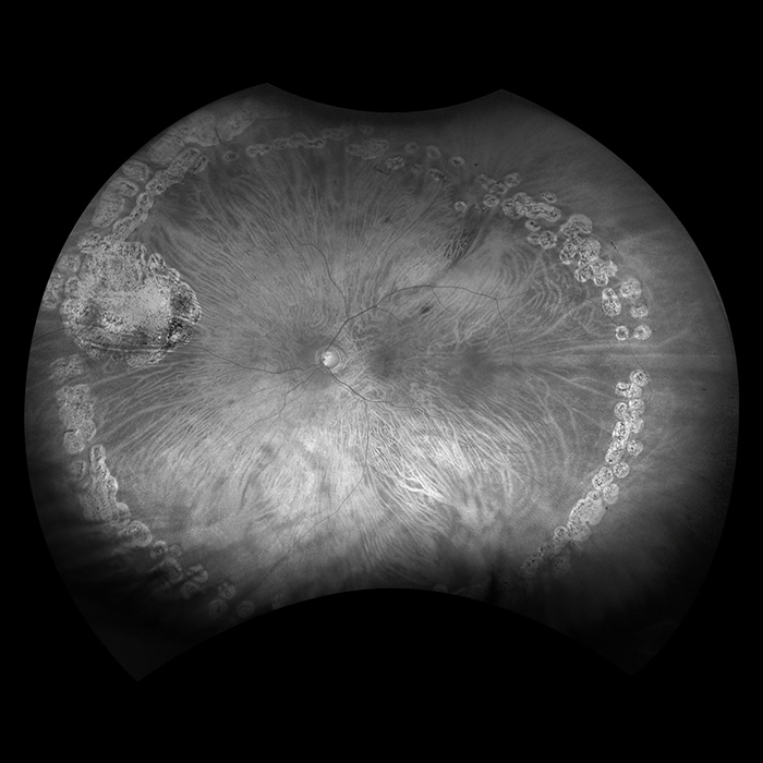
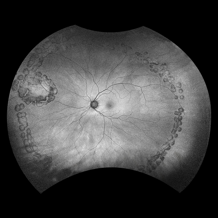
- Green and Blue Autofluorescence imaging to support analysis of retinal health
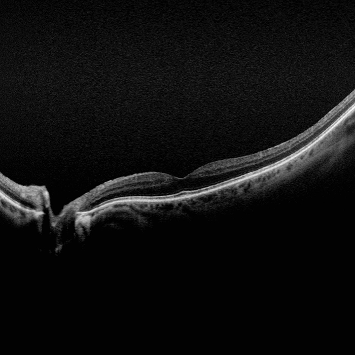
- Integrated high-resolution SD-OCT in every exam, elevating the standard of care
Software Analysis Features
- OptosAdvanceTM Image Management software streamlines image review and consultations
- A comprehensive Reference Database (RDB) enables results of OCT analysis to be shown in relation to 1%, 5%, 95%, and 99% of the RDB population
- Monaco RGB is equipped with AreaAssist, a tool designed to improve the efficiency of retinal imaging workflows by enabling users to automatically measure continuous areas of matching color and adjust the sensitivity of the selected area
- Distance (mm) and area (mm2) measurements provide objective assessment of change over time
Software Compliance Features
- Monaco RGB includes robust cybersecurity enhancements to comply with ISO27032 guidelines and the FDA Cybersecurity in Medical Devices* requirement, providing enhanced protection against threats
- DICOM-compatible software supports compliance with the Code of Federal Regulations3
1 Legarreta. Imaging of Peripheral Retina with Optos Ultra-Widefield Imaging: Evaluation of Aperture Size on Image Quality. ARVO 2012.
2 Friberg. Advances in retinal imaging of eyes with hazy media: Further Studies. ARVO 2011.
3 All Covered Entities must securely backup ‘retrievable exact copies of ePHI’ (CFR 164.308 (7)(ii)(A)).
*For US markets only
**Monaco RGB holds a CE mark contact us for availability
Technical Specifications
| Image Modalities |
optomap color rgb (red, green, blue laser) |
|---|---|
| Resolution |
optomap plus: 14 μm |
| Exposure Time |
Less than 0.4 seconds |
| OCT Scan Types |
Line Scan |
| Wavelengths |
Red laser: 635 nm |
| Tomographic Imaging |
Signal Type: Optical scattering from tissue |
| OCT Scan Characteristics |
Spectral Domain OCT |
| Footprint |
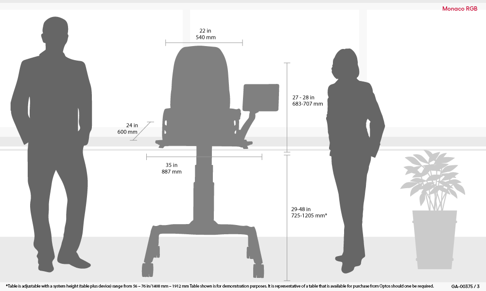
Width: 540 mm/22 inches |
Image Modalities

Monaco RGB Device Footprint