optomap Color RGB: A More Natural Ultra-widefield View
optomap color RGB imaging offers eye care professionals an advanced ultra-widefield (UWF™) view designed to enhance the assessment of retinal, choroidal, and optic nerve conditions. By capturing detailed images with multiple laser wavelengths, this innovative technology provides a clearer, more natural perspective of the eye, supporting earlier detection, improved diagnostic accuracy, and greater clinical confidence. Backed by extensive research and thousands of peer-reviewed publications, optomap continues to set the standard in retinal imaging and helps practitioners deliver better patient care.
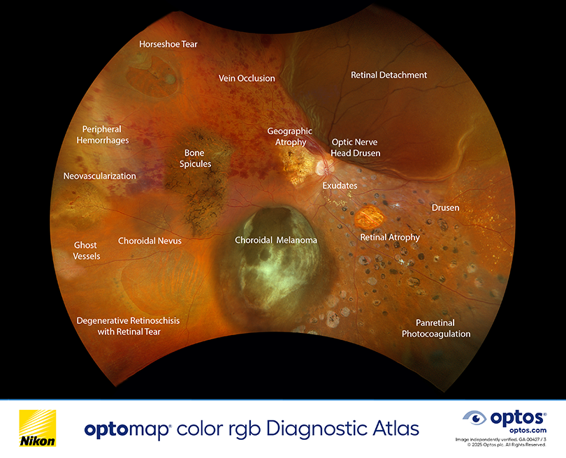
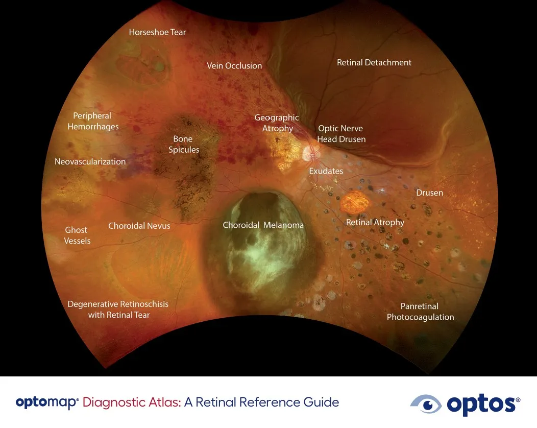
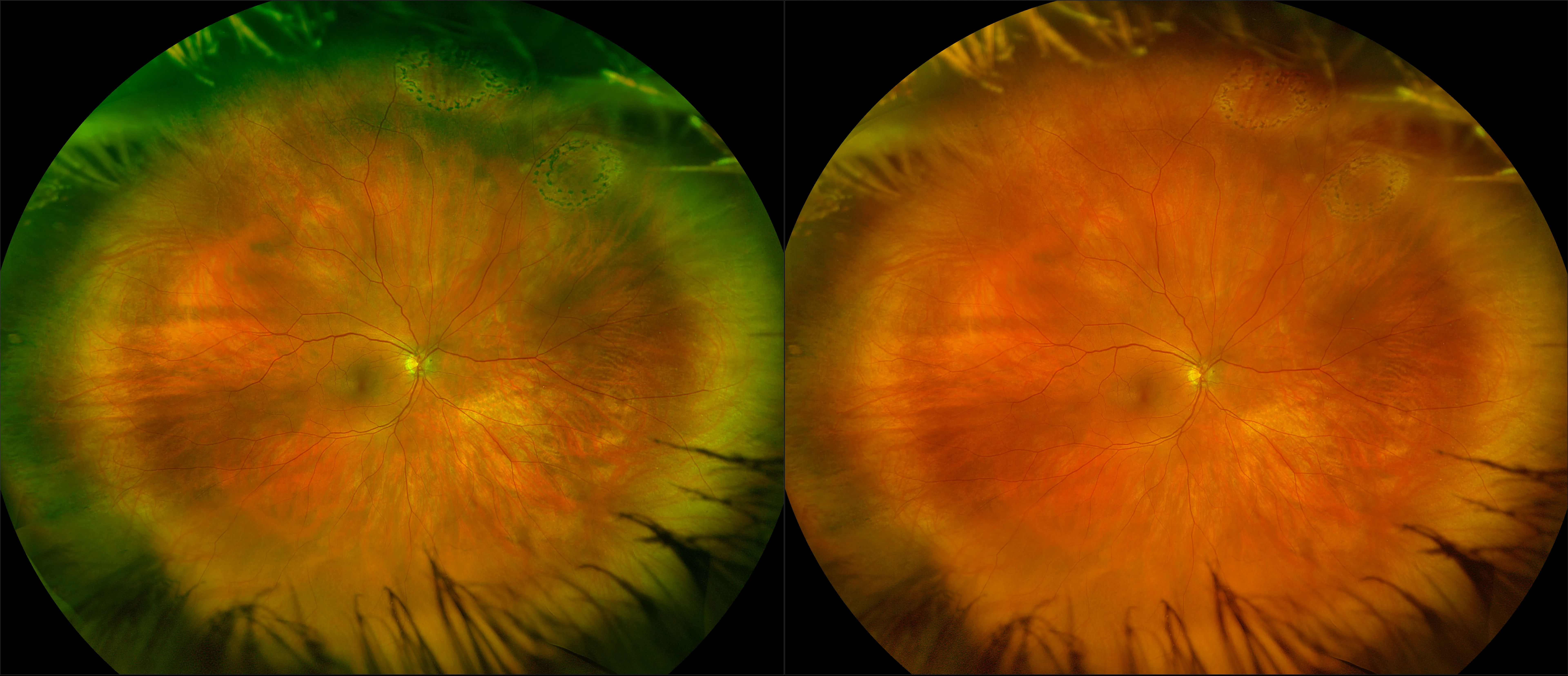
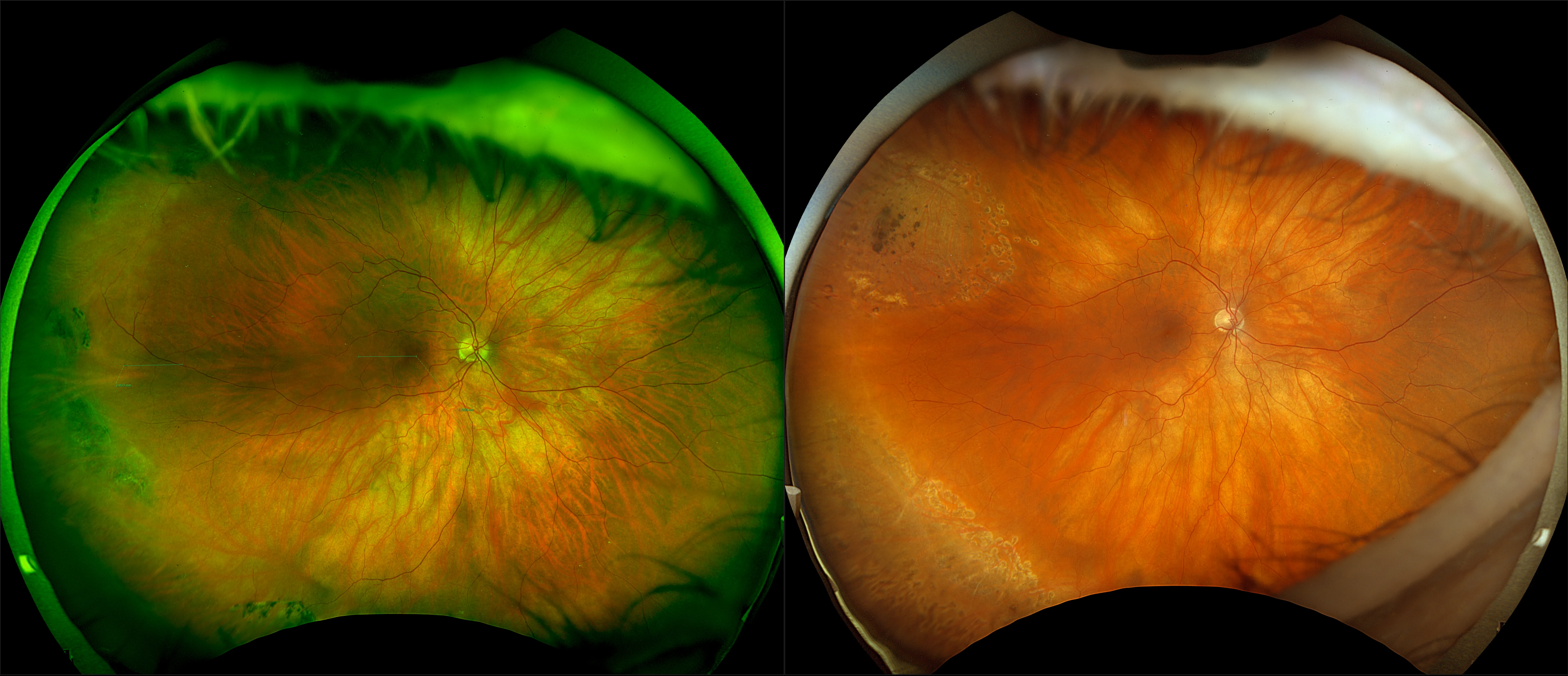
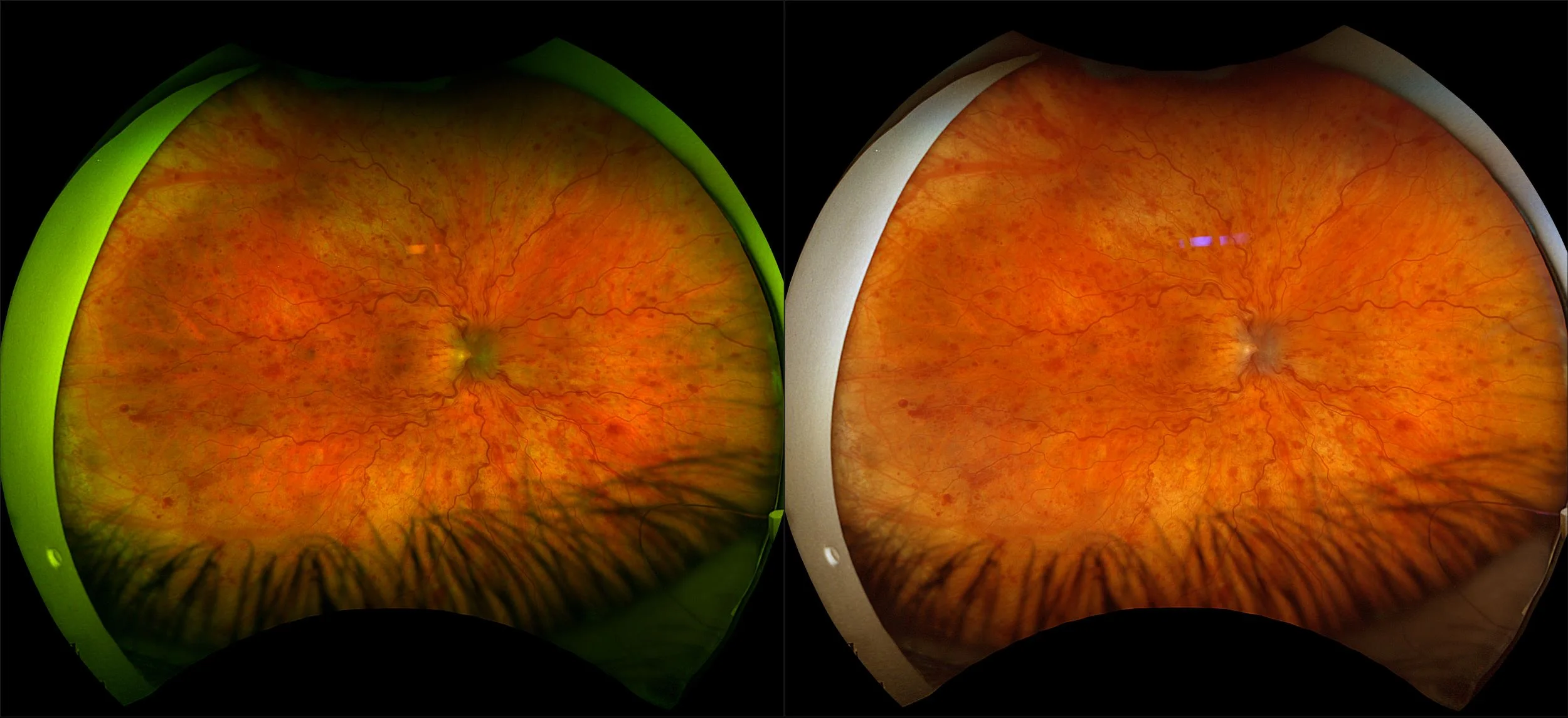
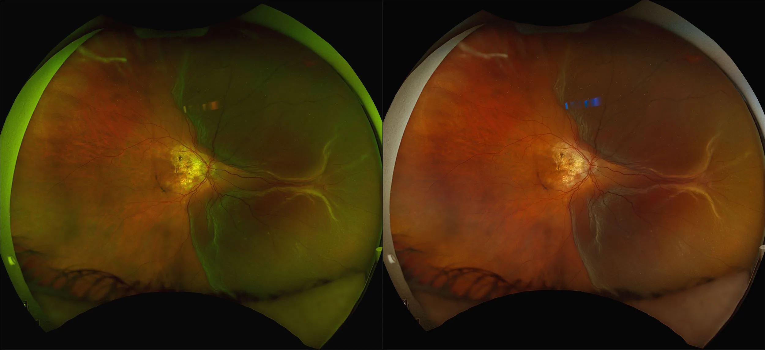
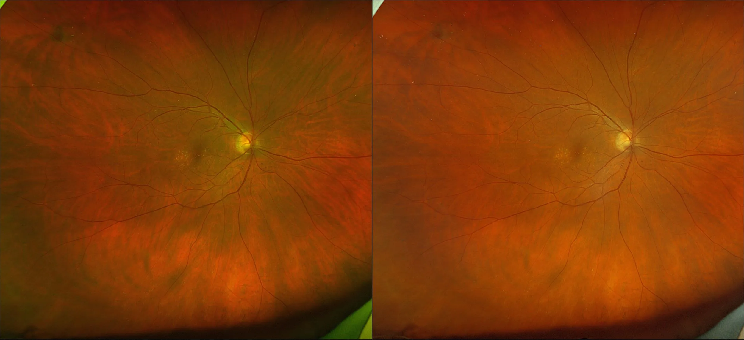
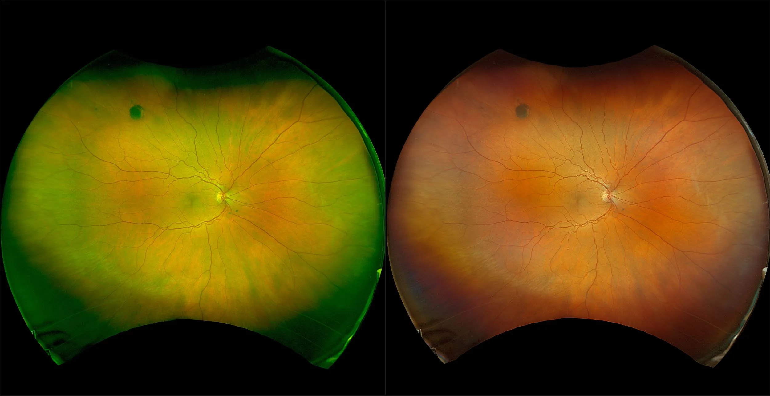
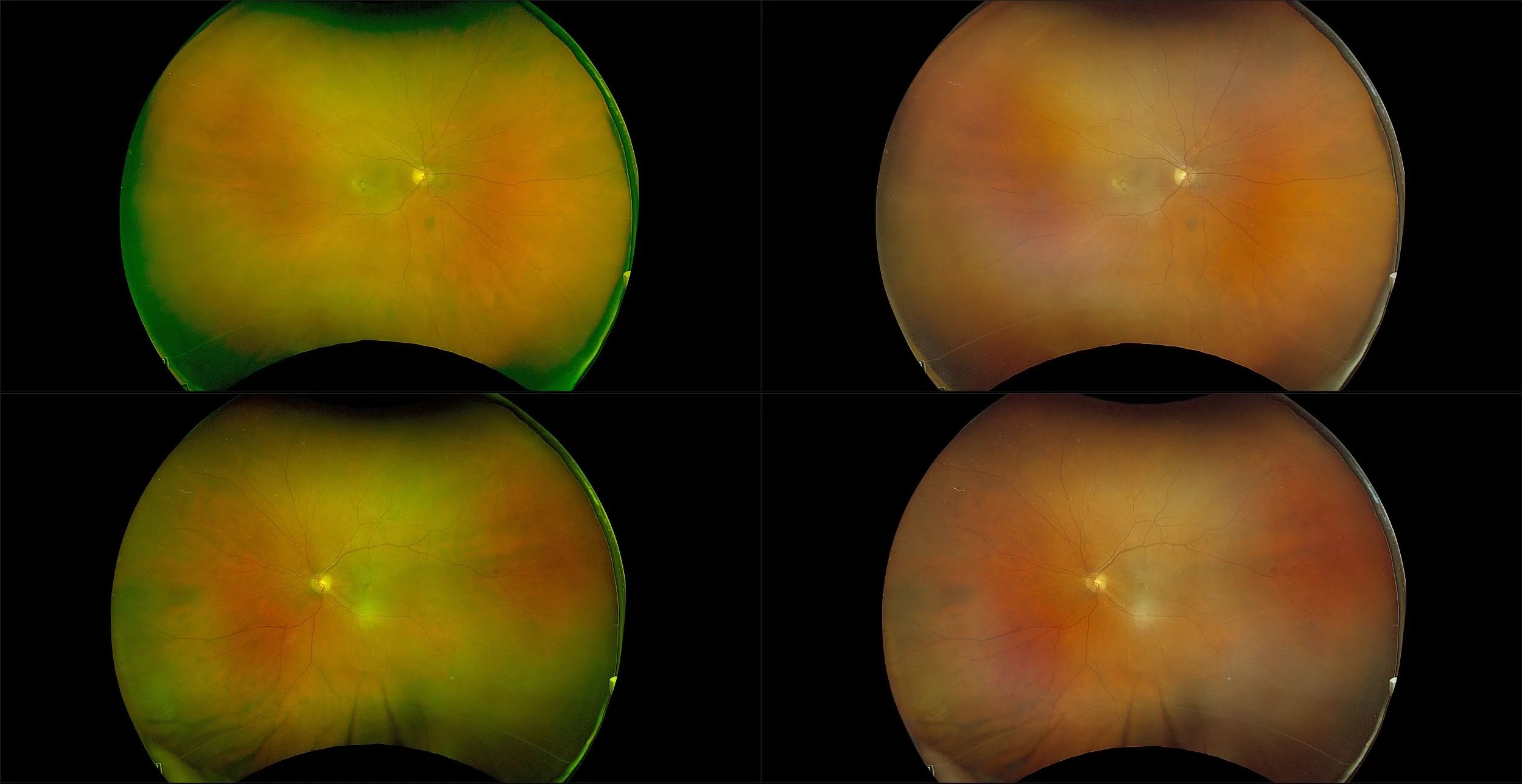
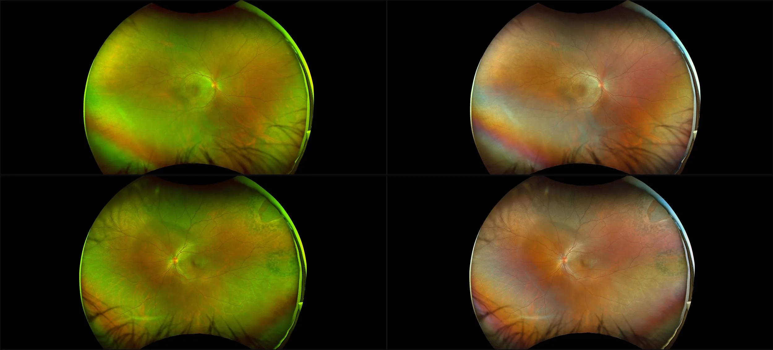
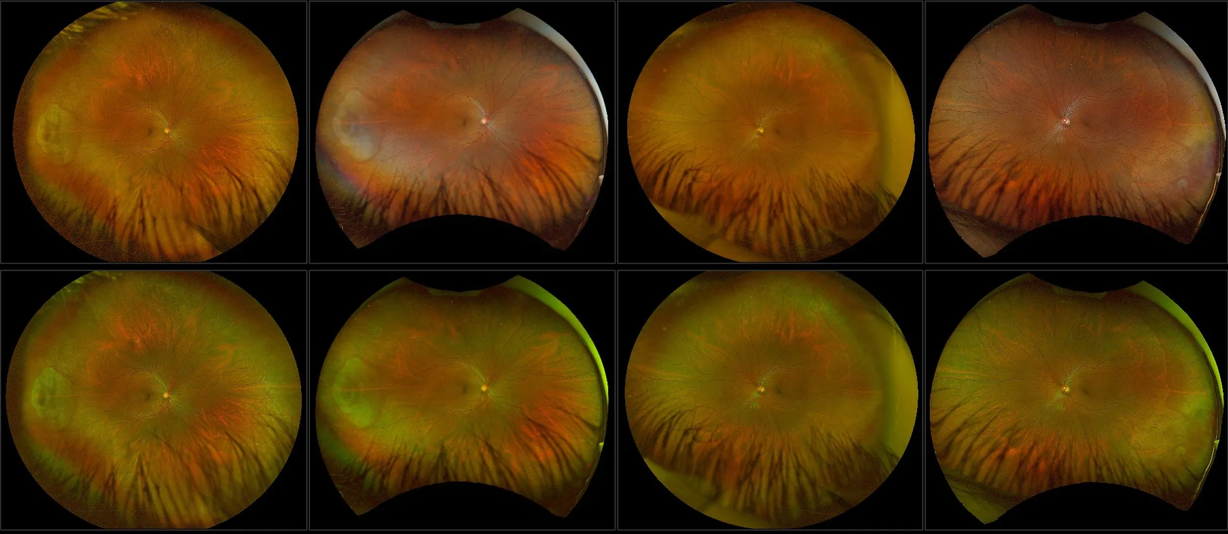
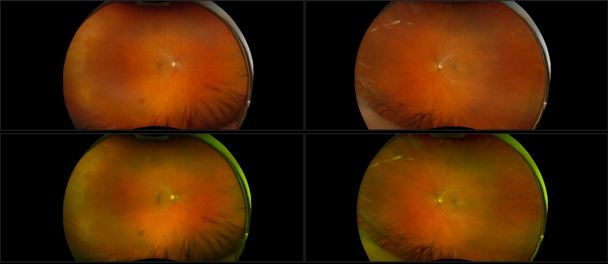
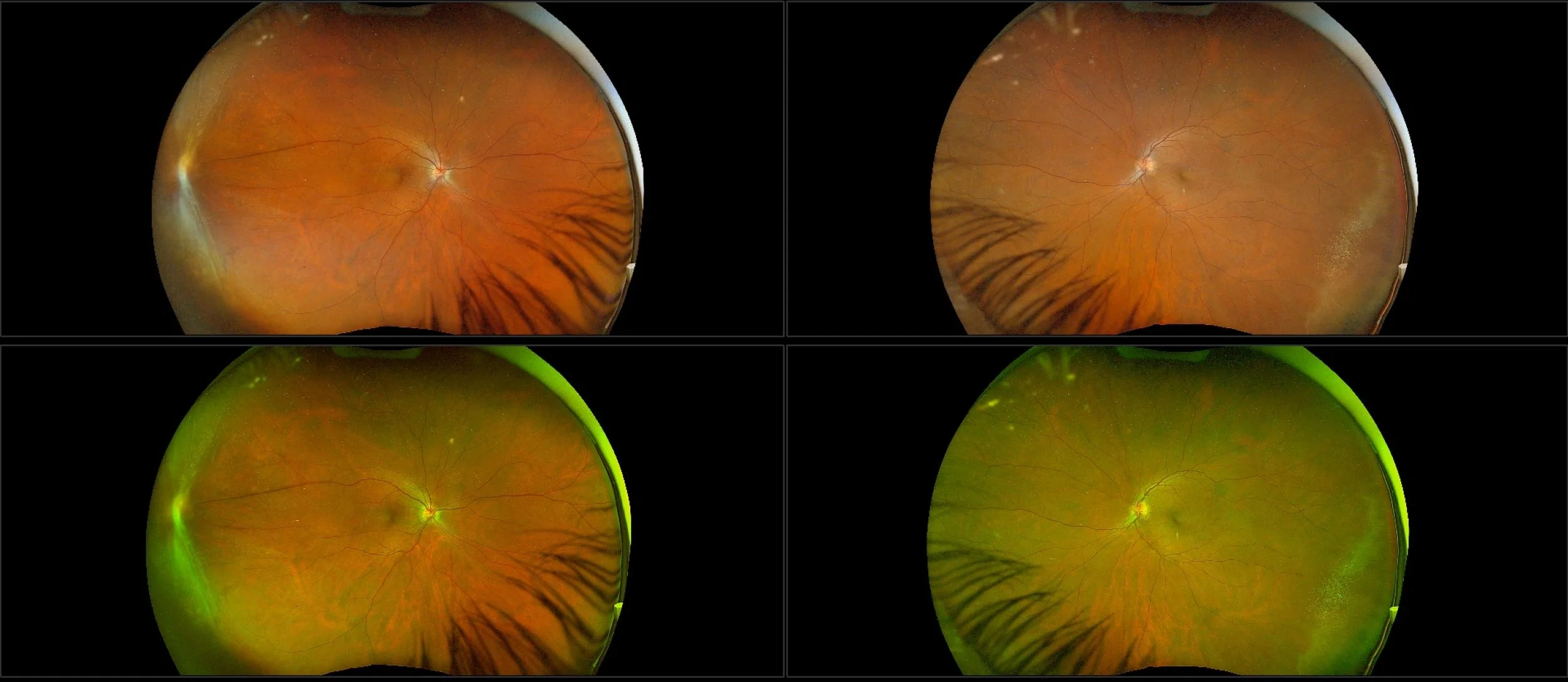
optomap Color RGB Confers Advantage
optomap color rg has long been the standard for UWF imaging, with more than 4,000 peer-reviewed publications underlining its utility for diagnosing, managing, and treating retinal disease. Color rg is the standard that Optos UWF imaging was based.
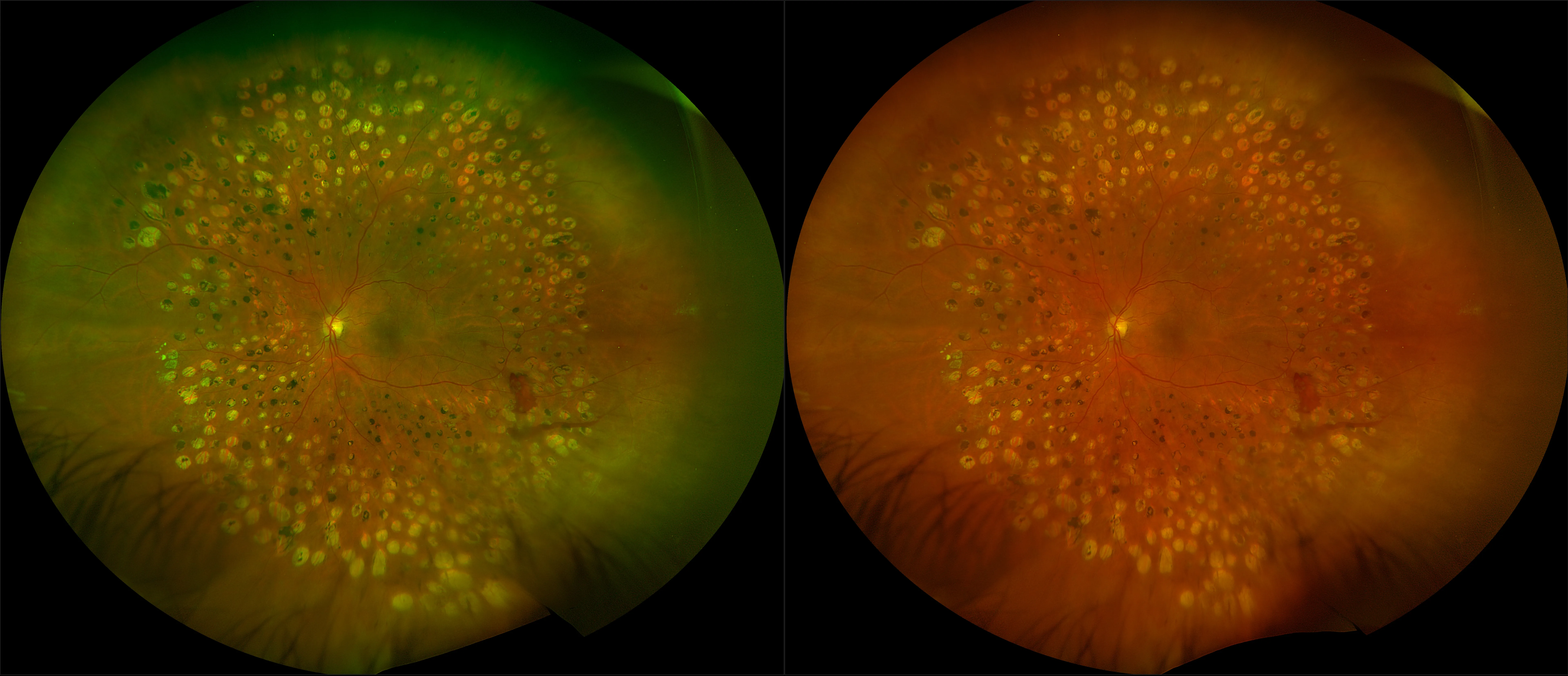
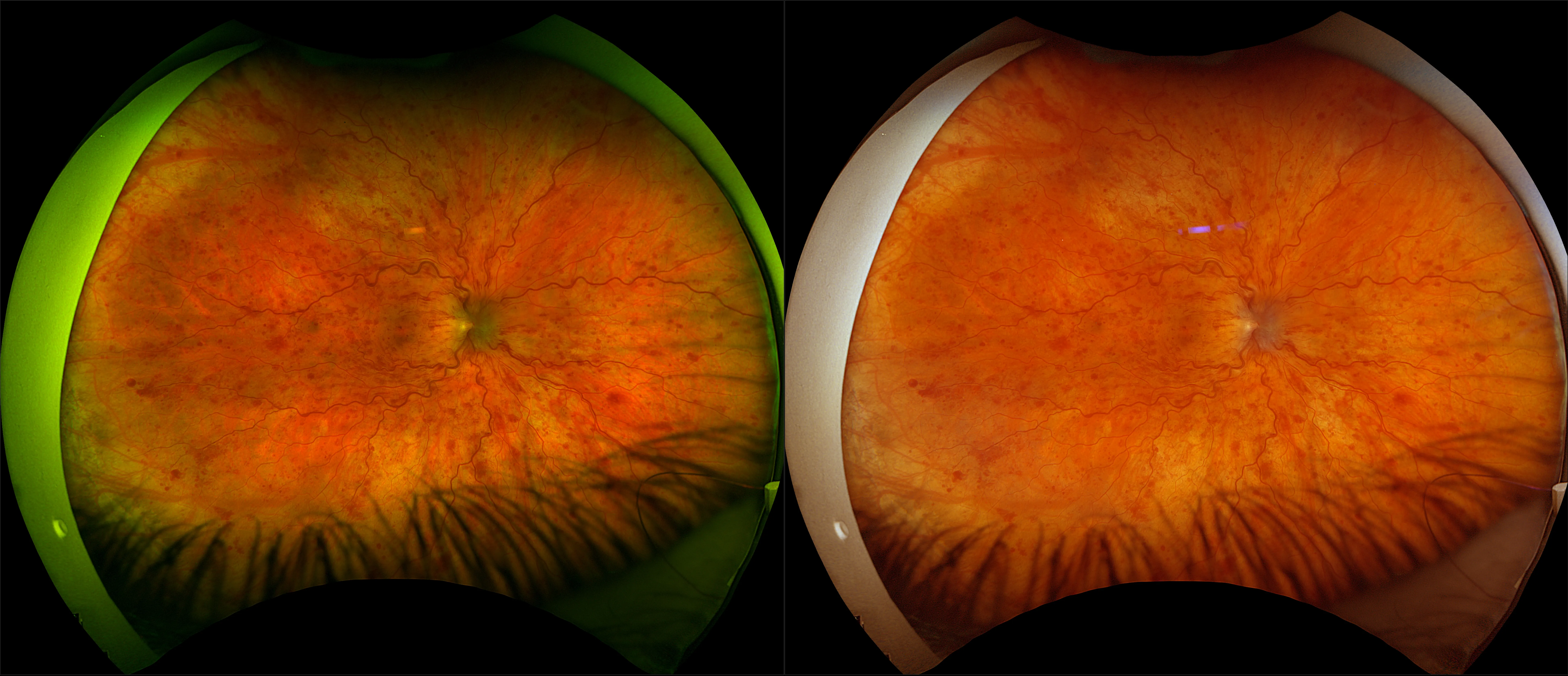
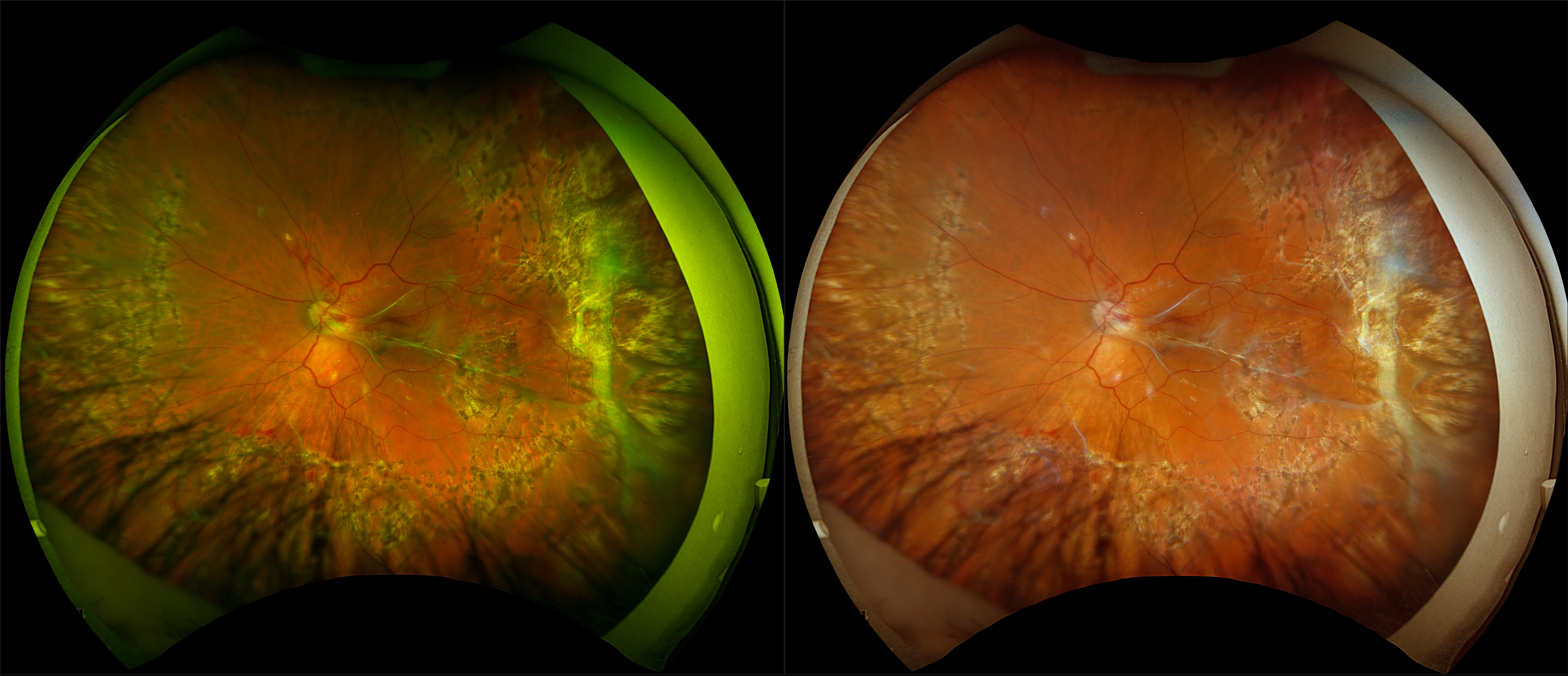
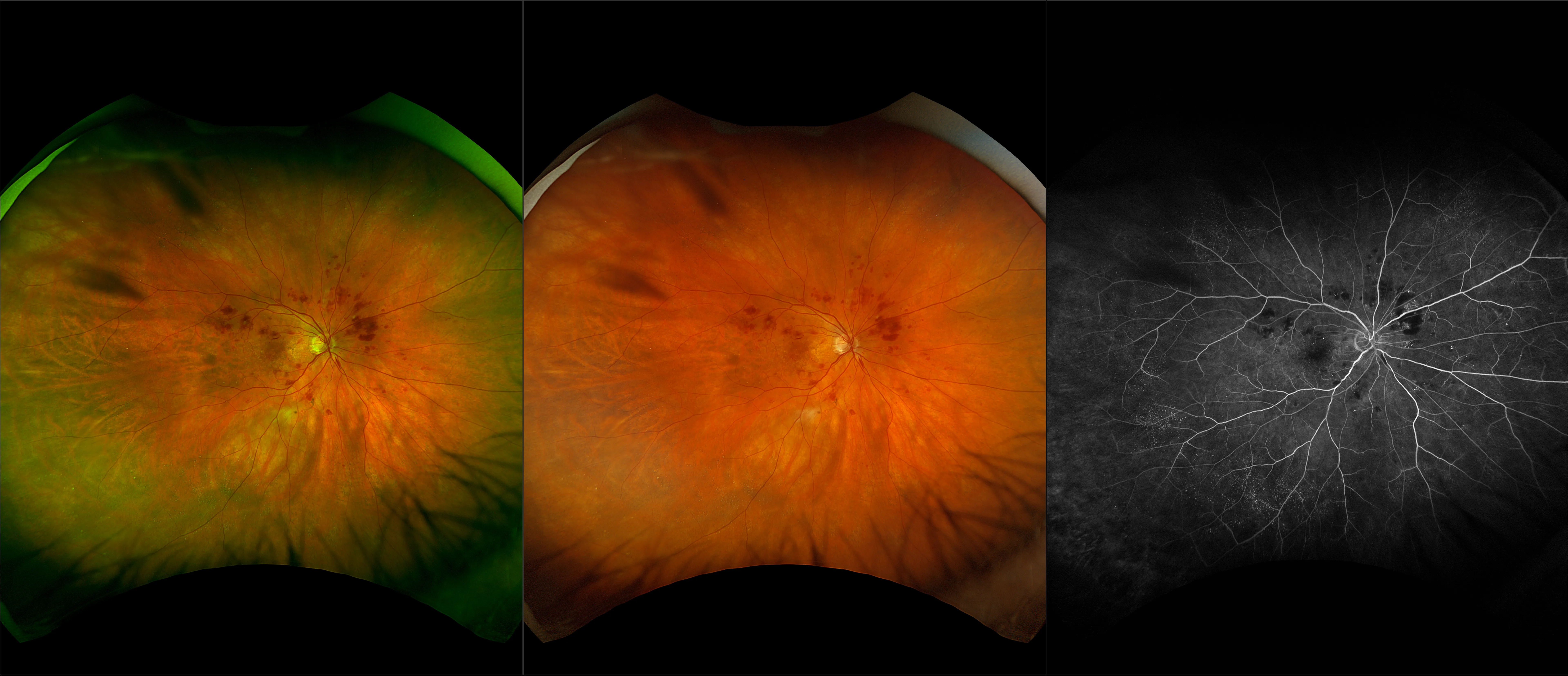
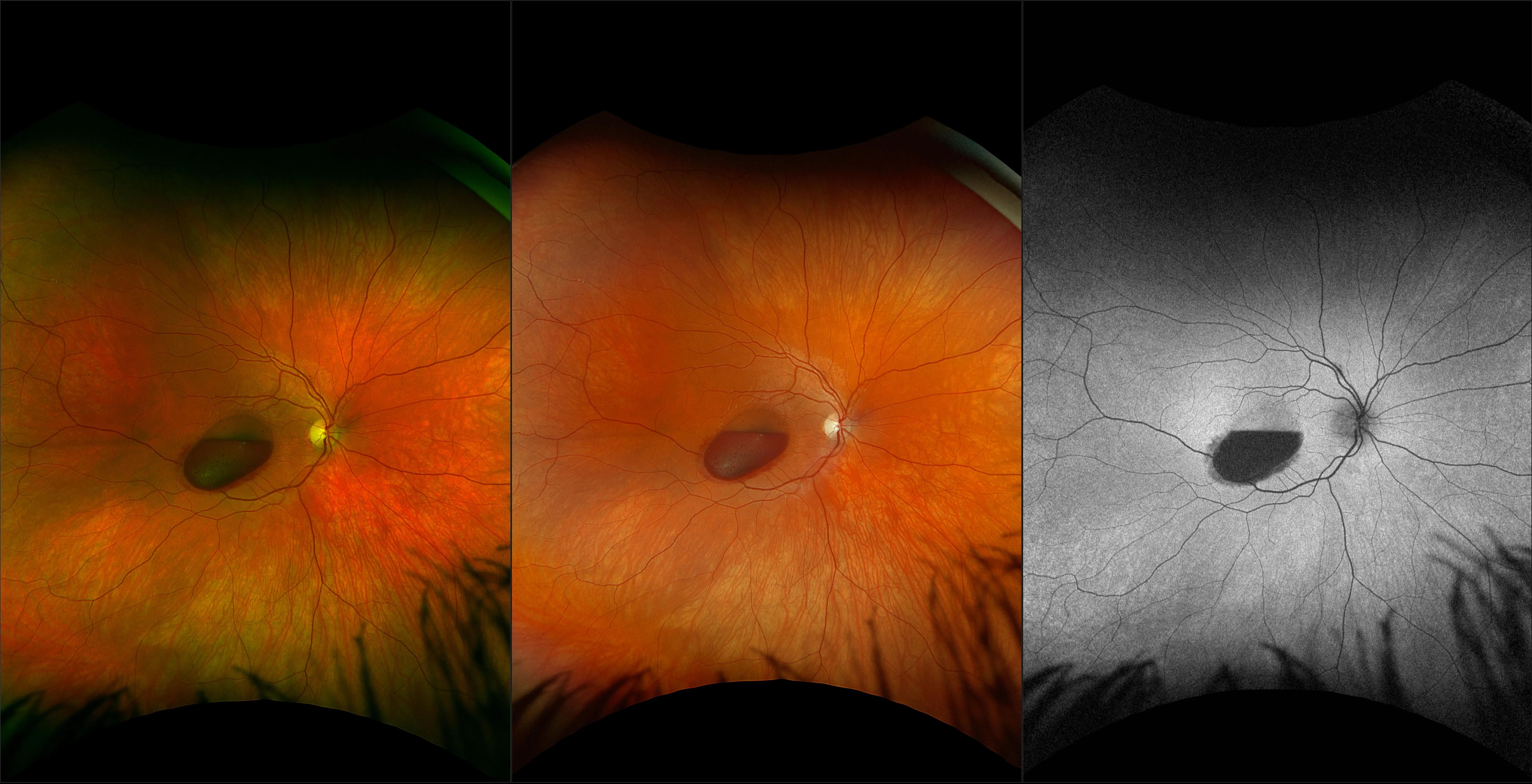
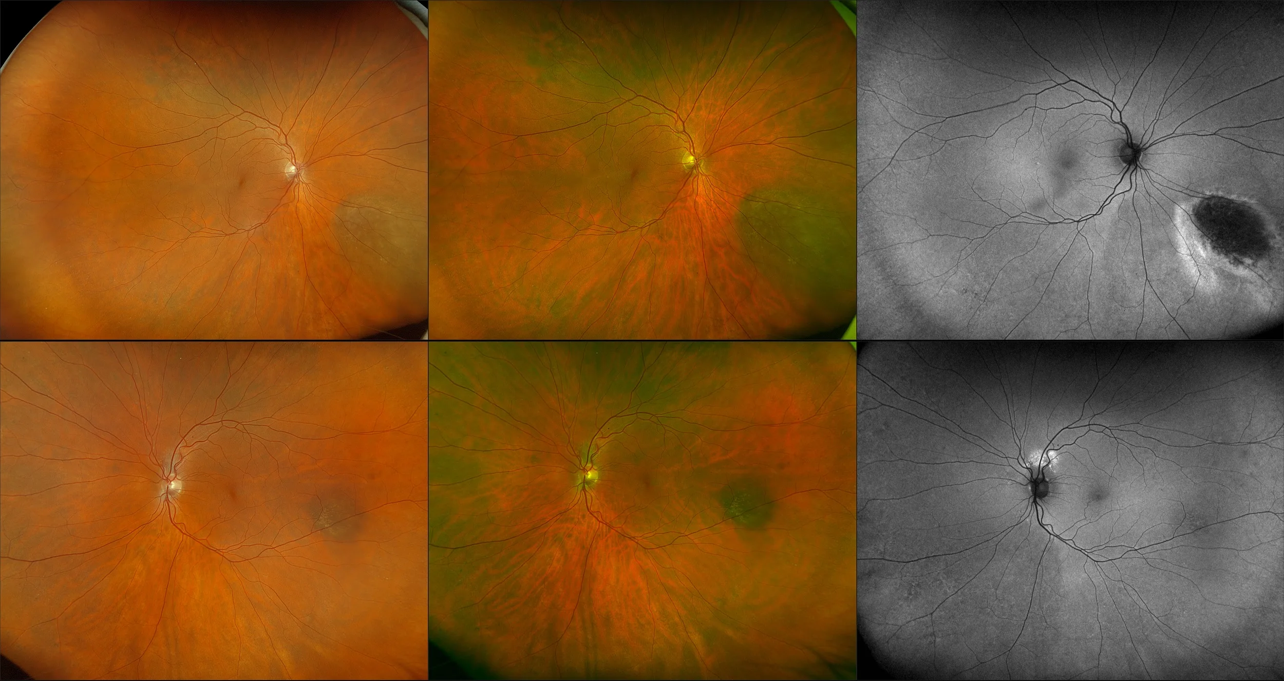
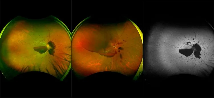
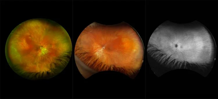
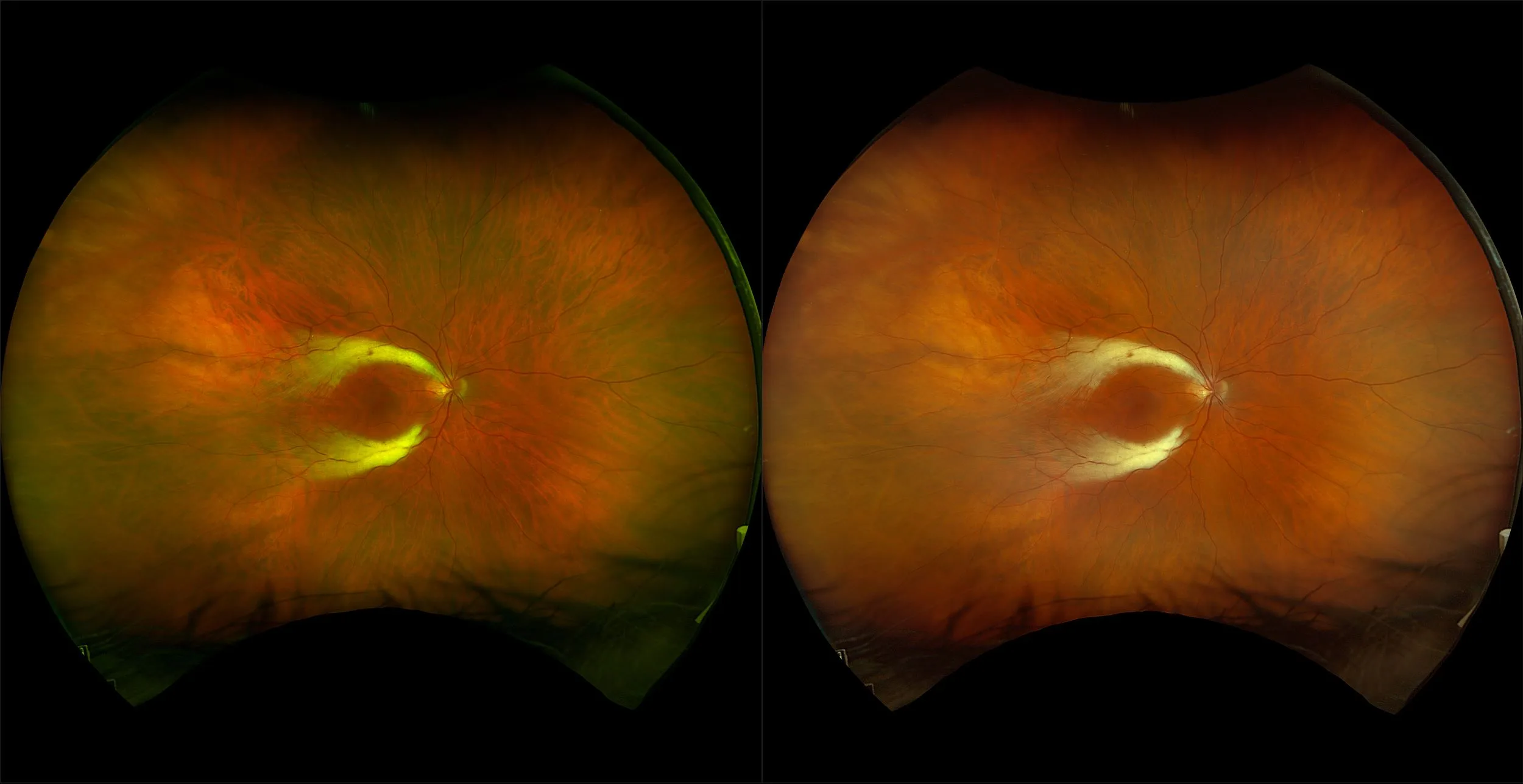
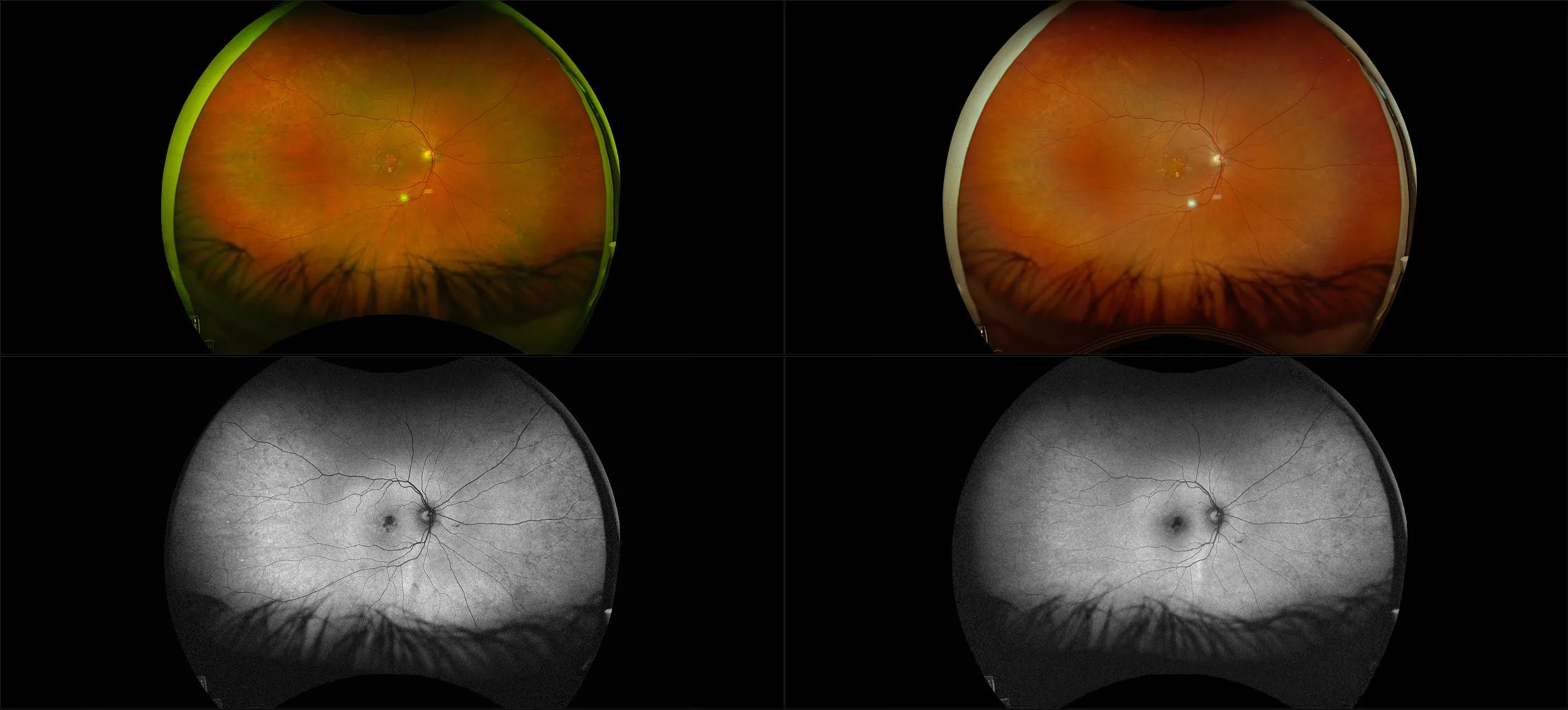
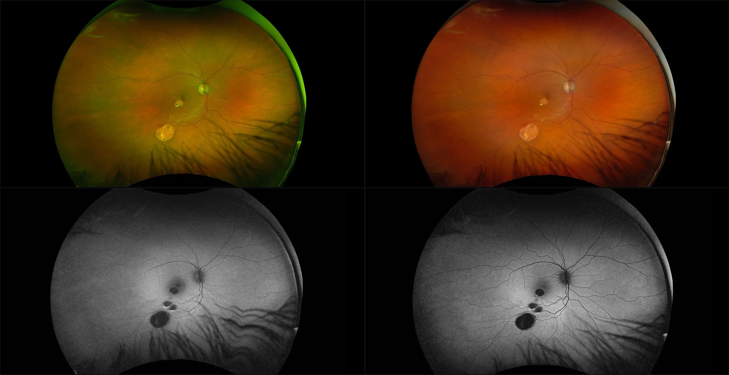
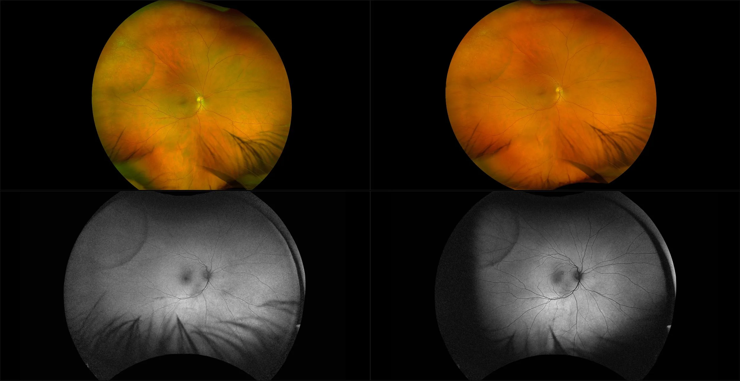
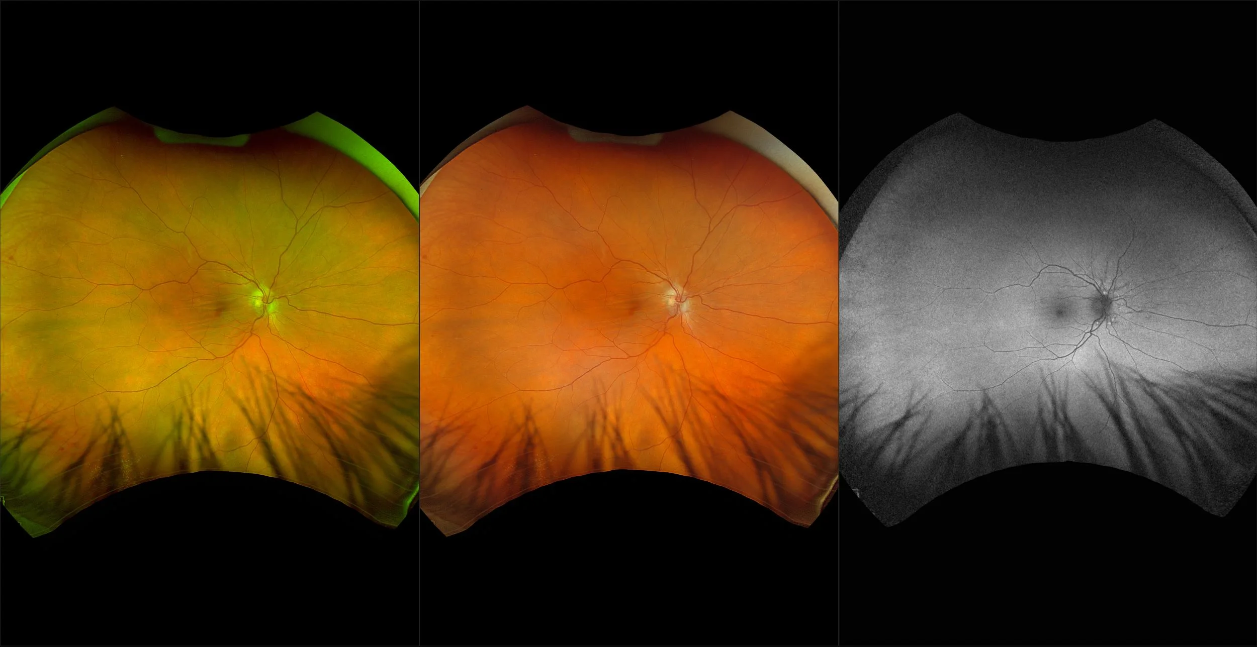
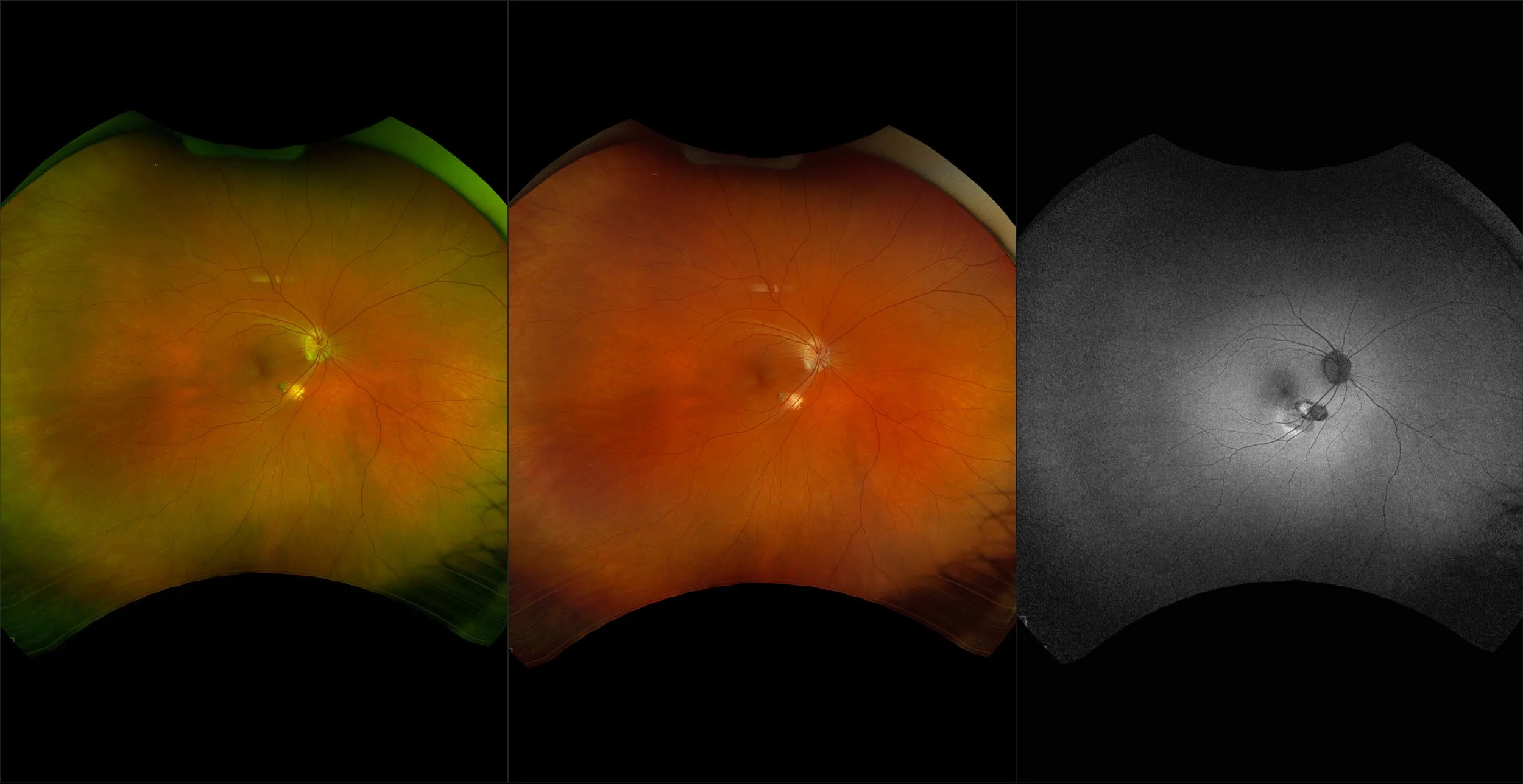
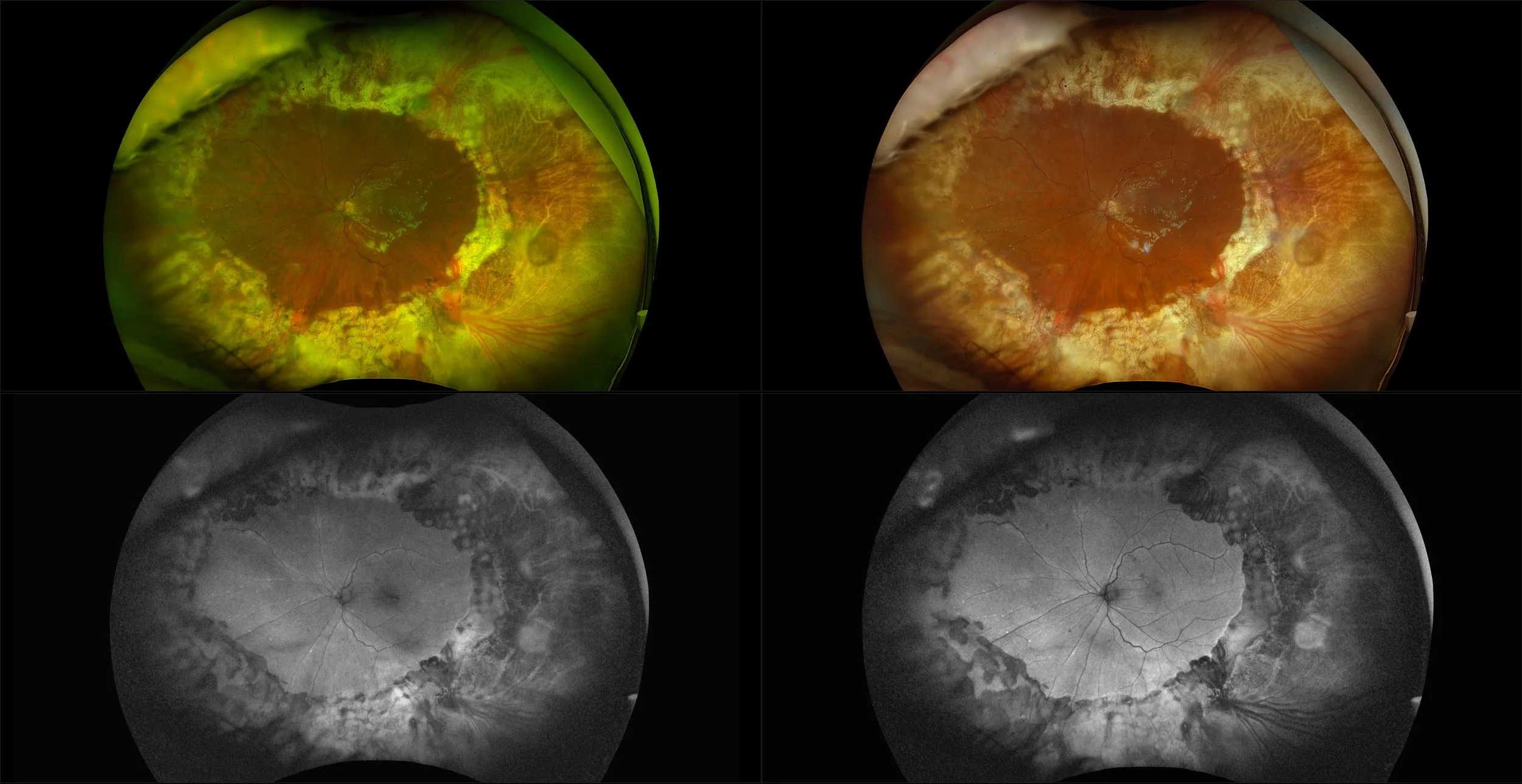
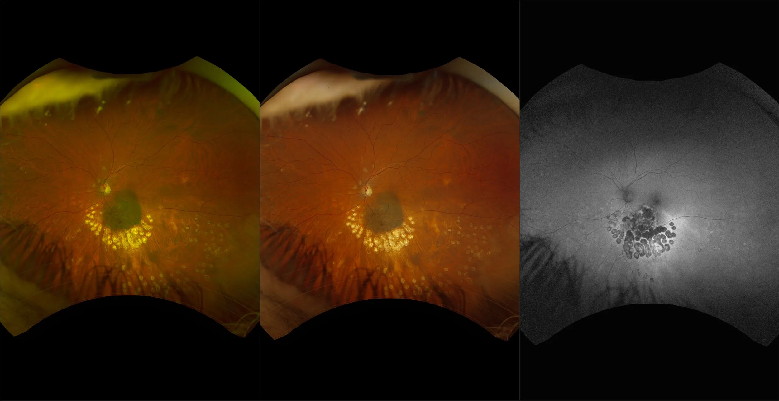
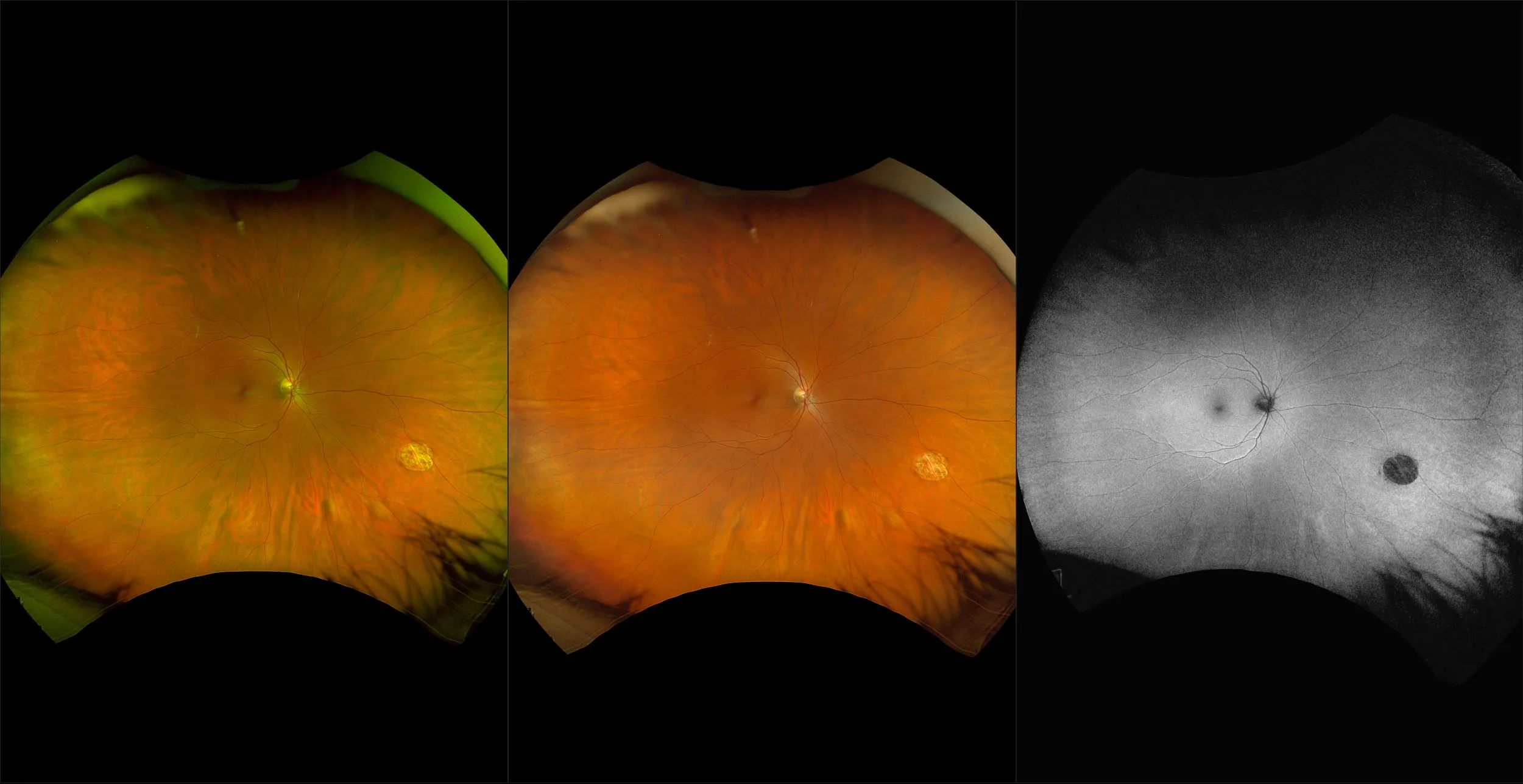
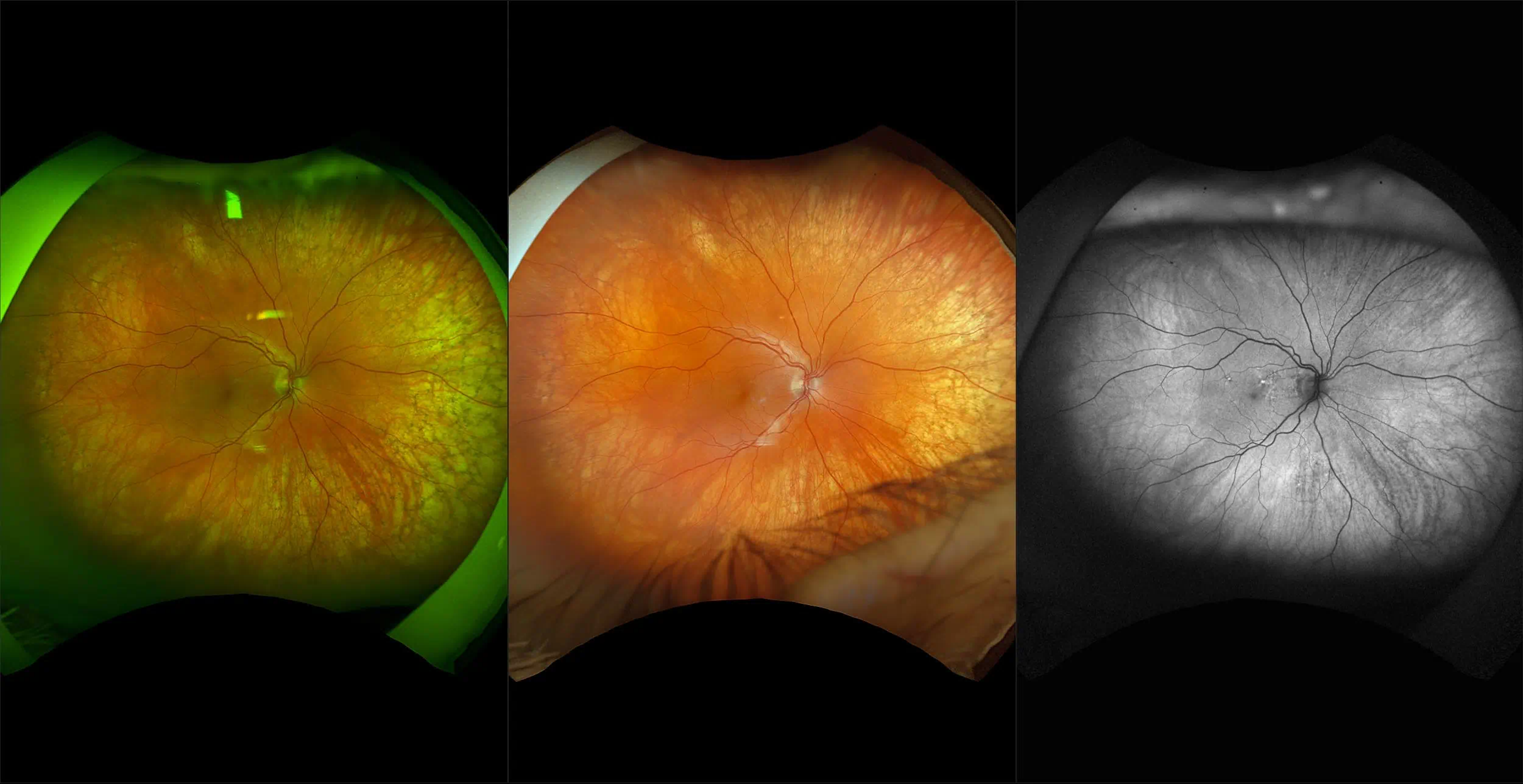
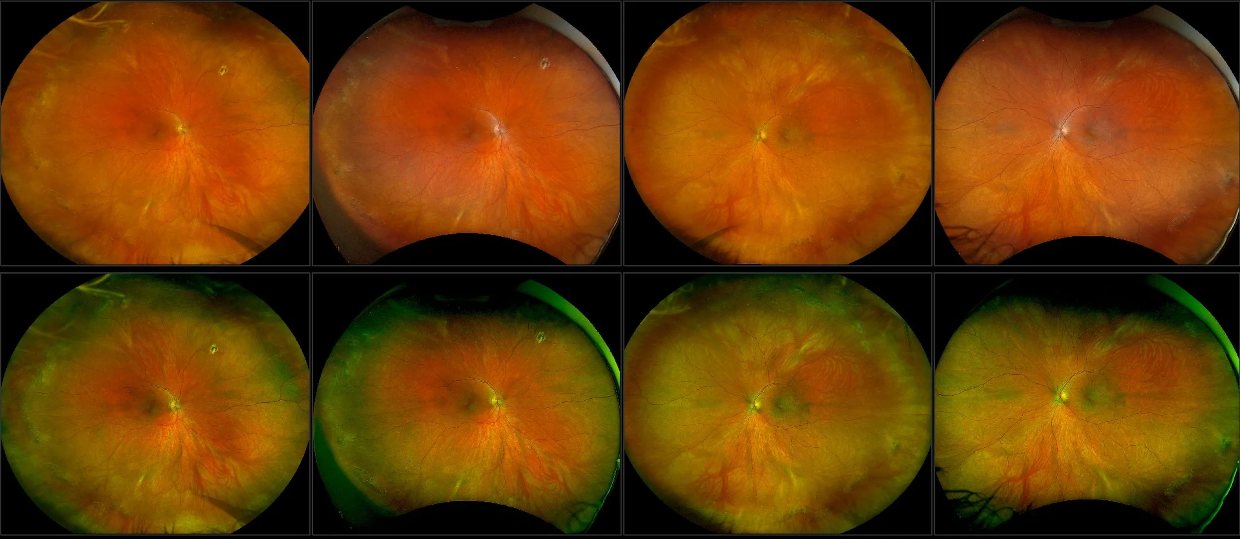
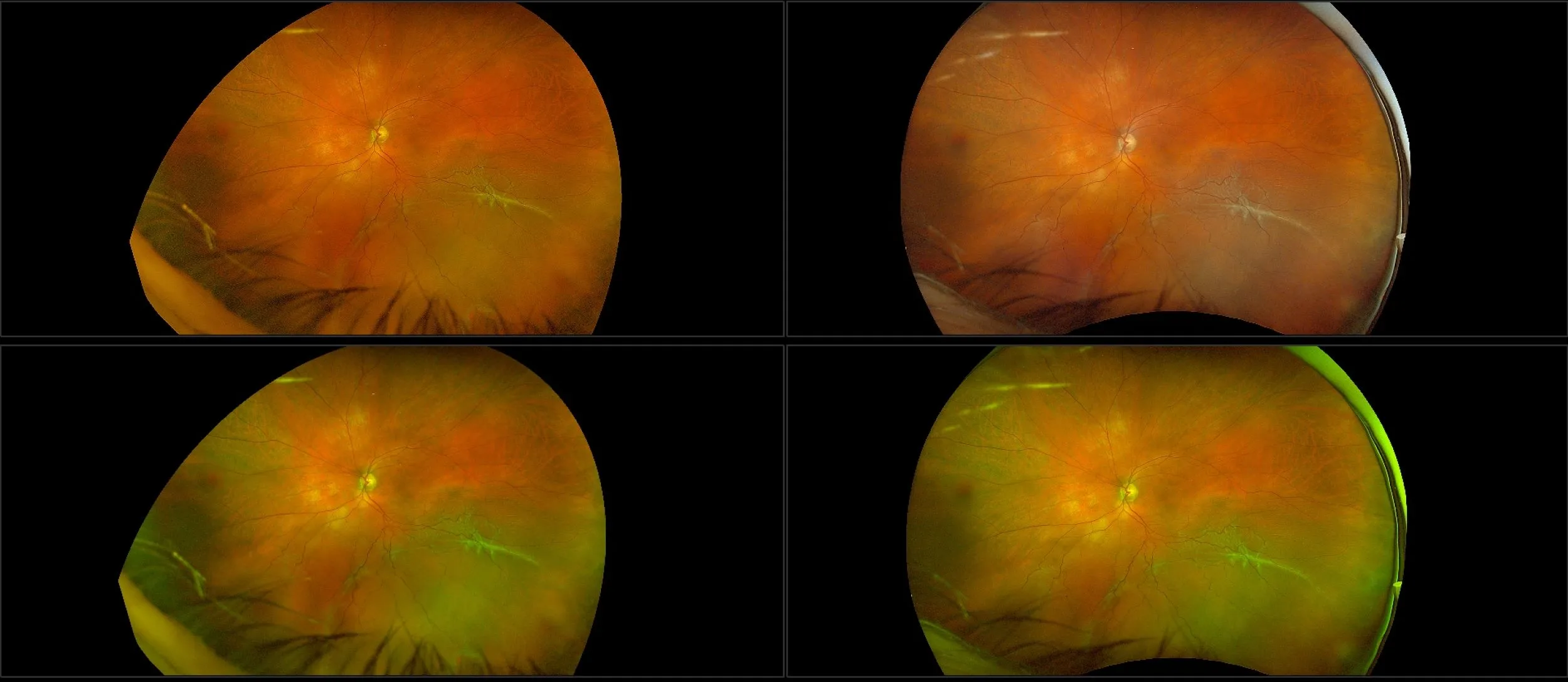
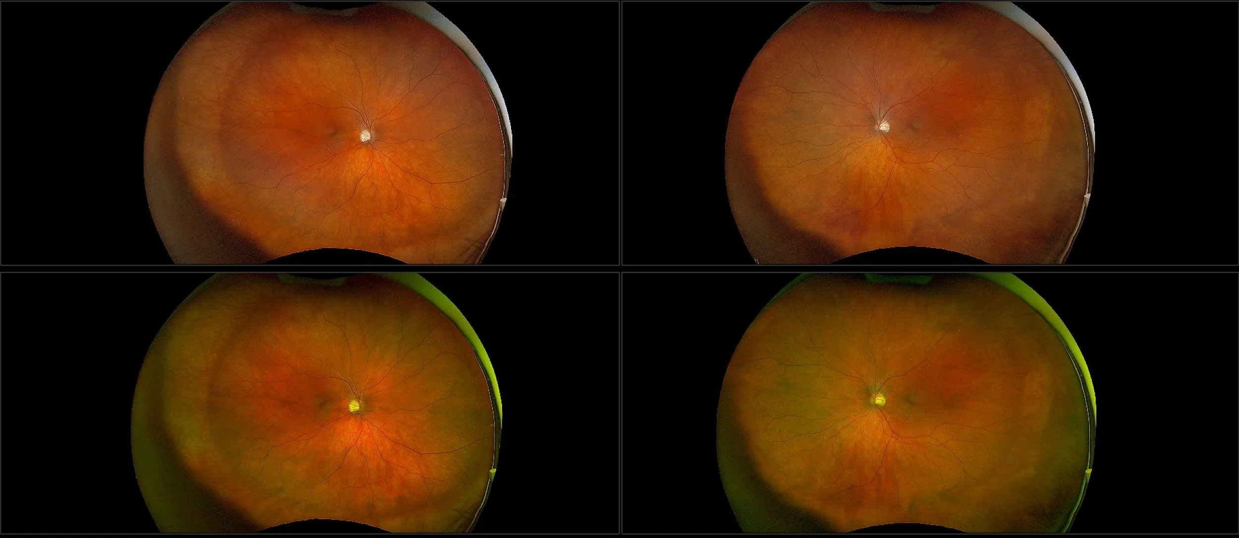
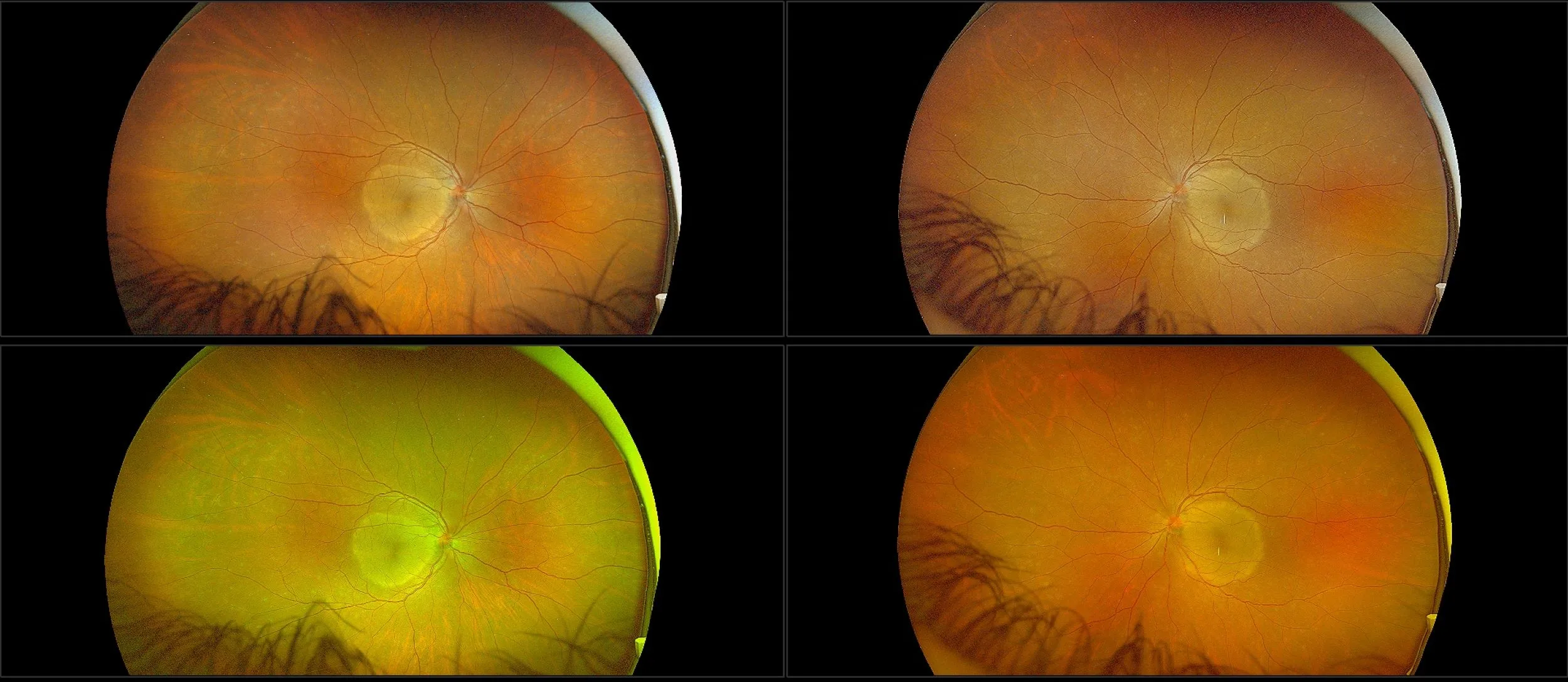
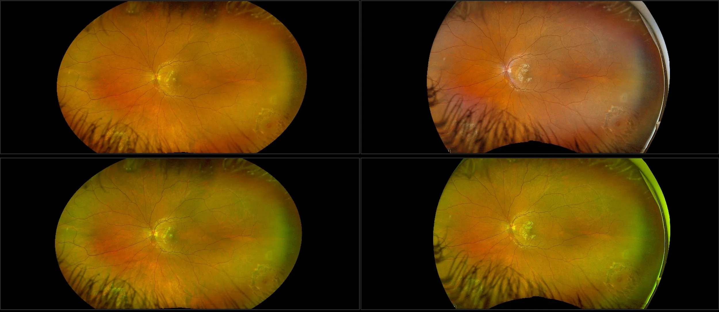
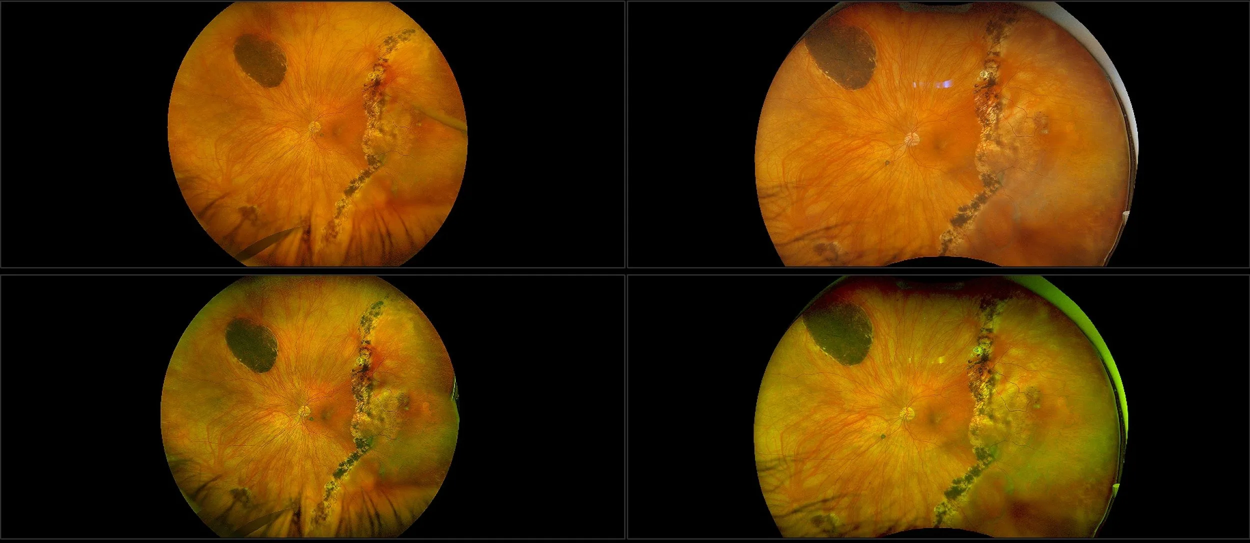
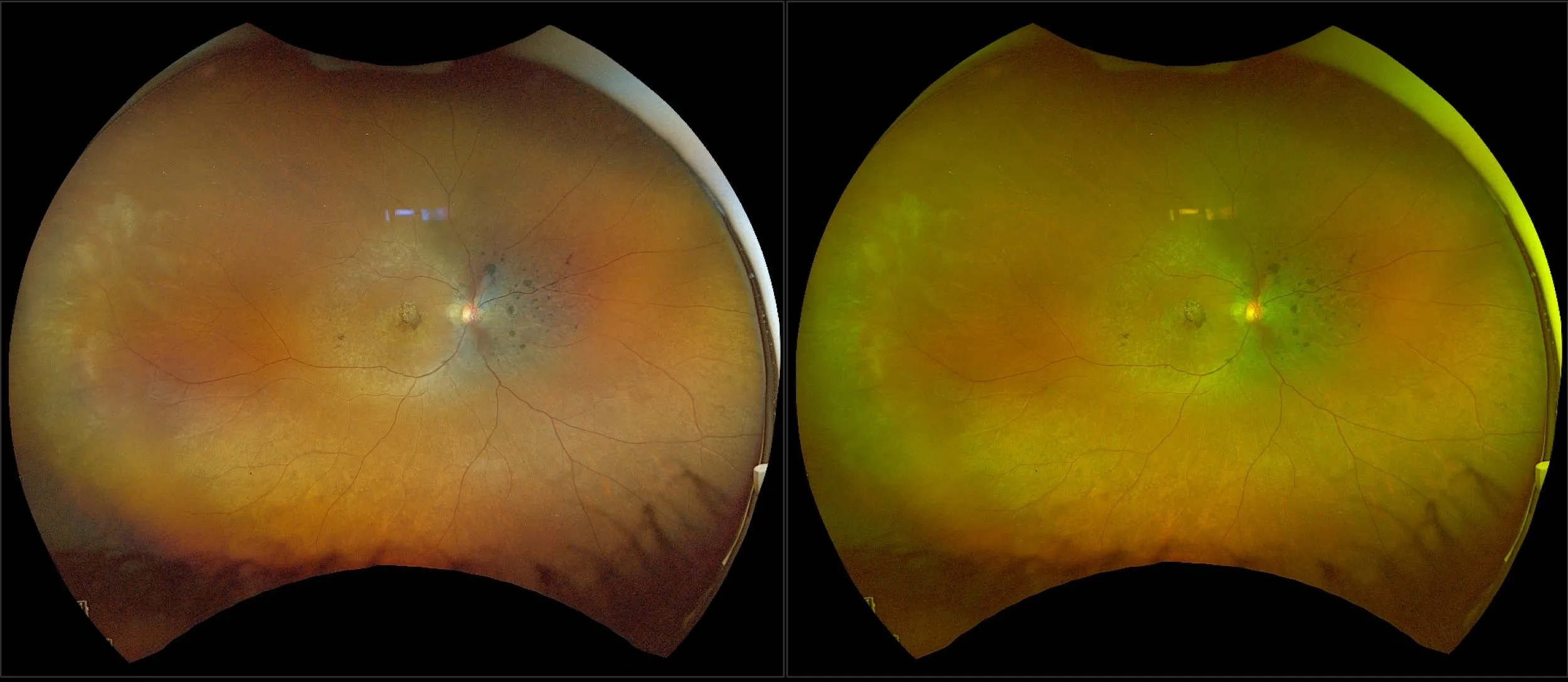
With its own merits optomap color rgb with four channel image review not only provides a more natural view of the retina but appears to confer an advantage for the following conditions:
- Optic nerve anatomy3
- Hyaloid reflection3
- PVR subretinal band3
- Superficial retinal hemorrhages3
- Neovascularization3
- Peripheral retinal abnormalities (holes, tears, lattice)3
- Ghost vessels or ischemia3
- Enhanced contrast between the retinopexy3
- Retinoschisis4
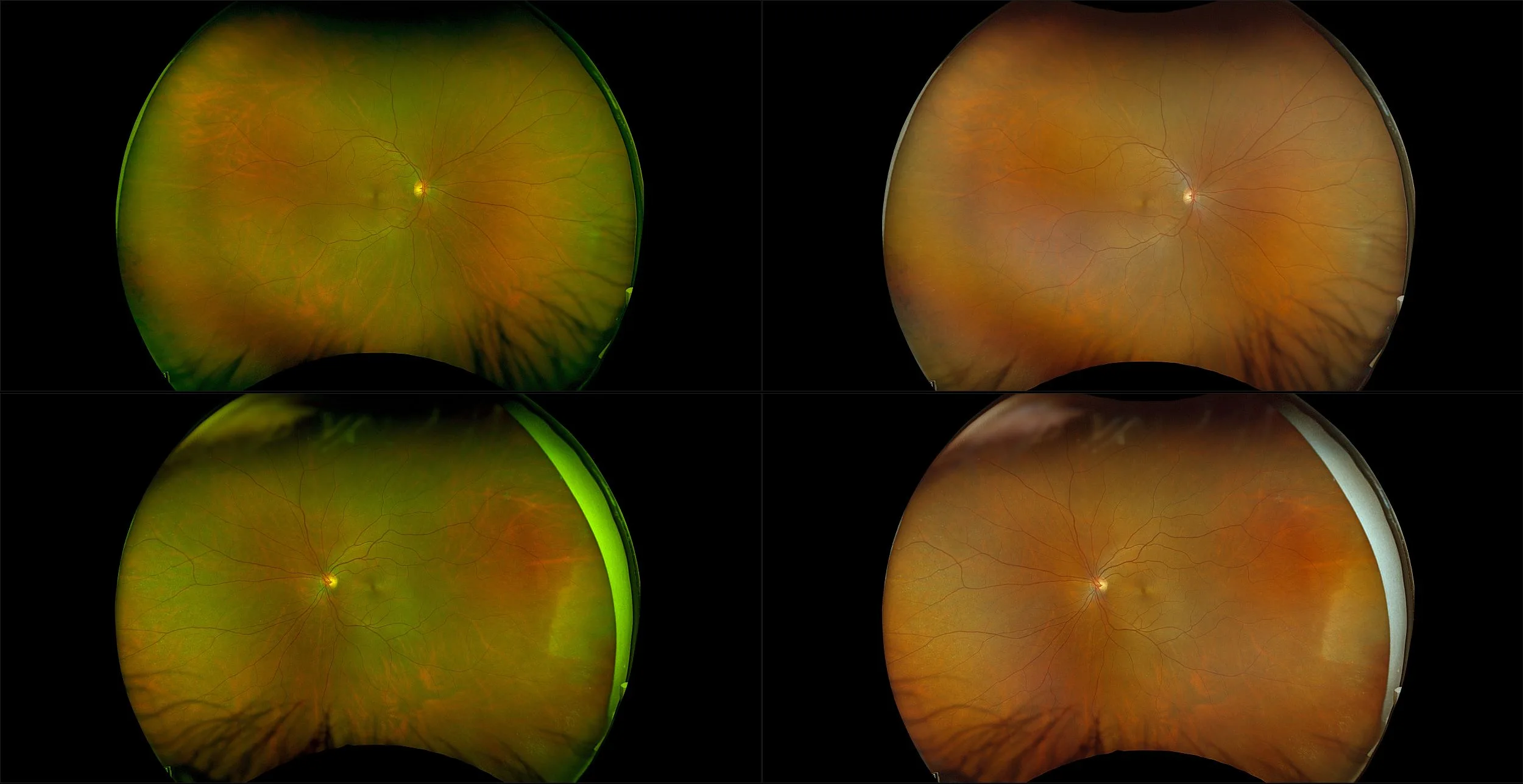
optomap Color RGB vs the Competition
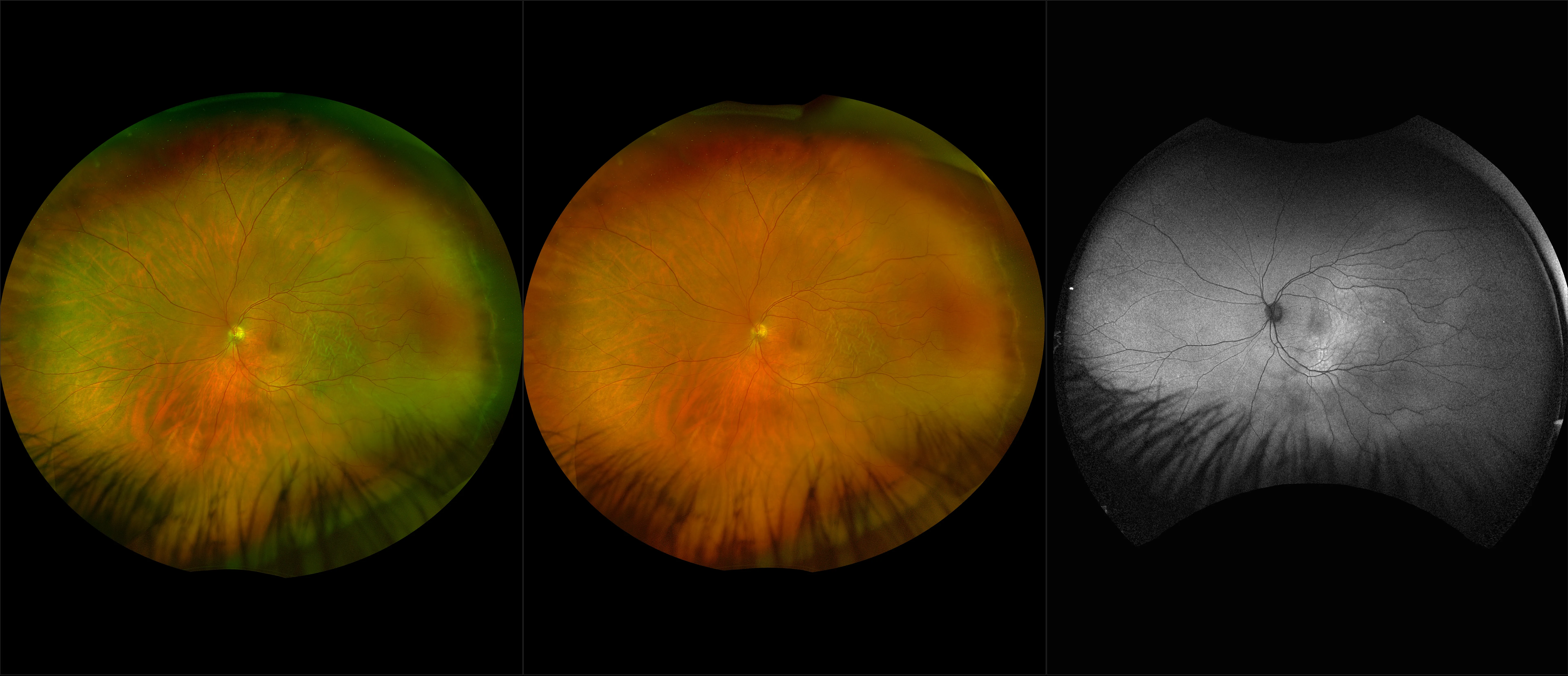
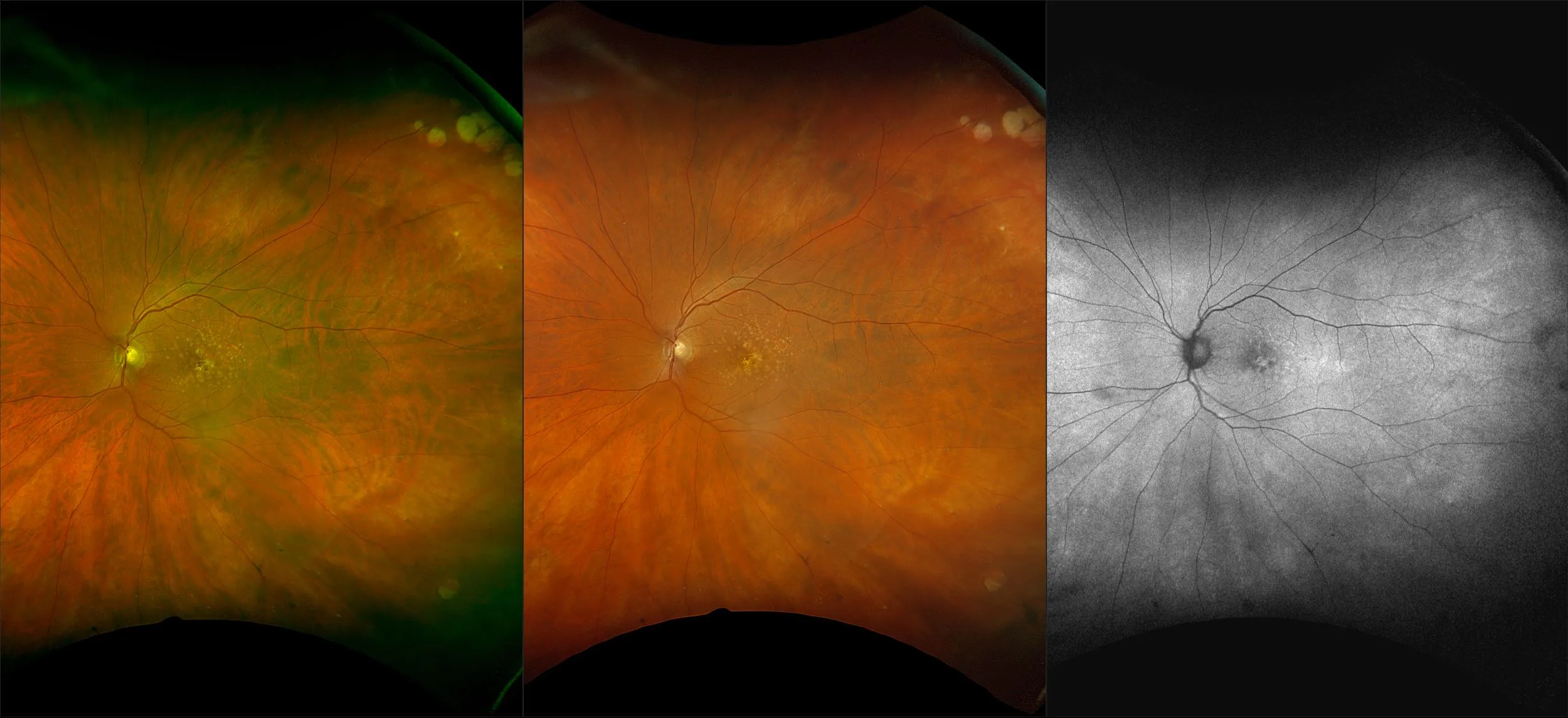
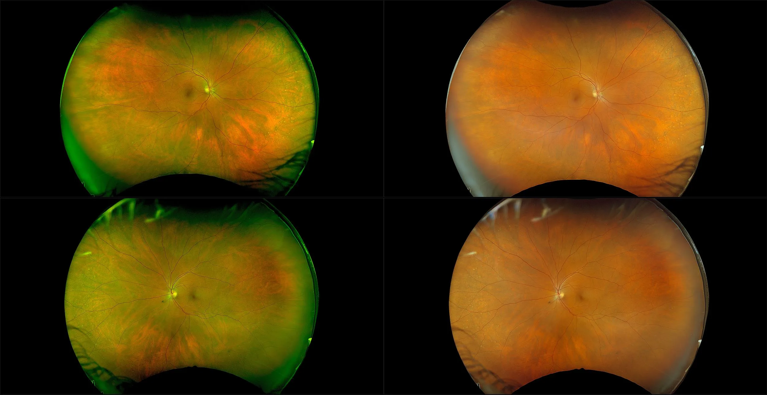
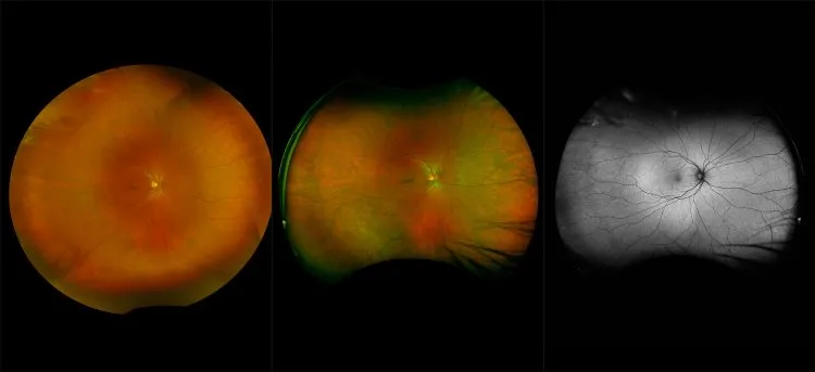
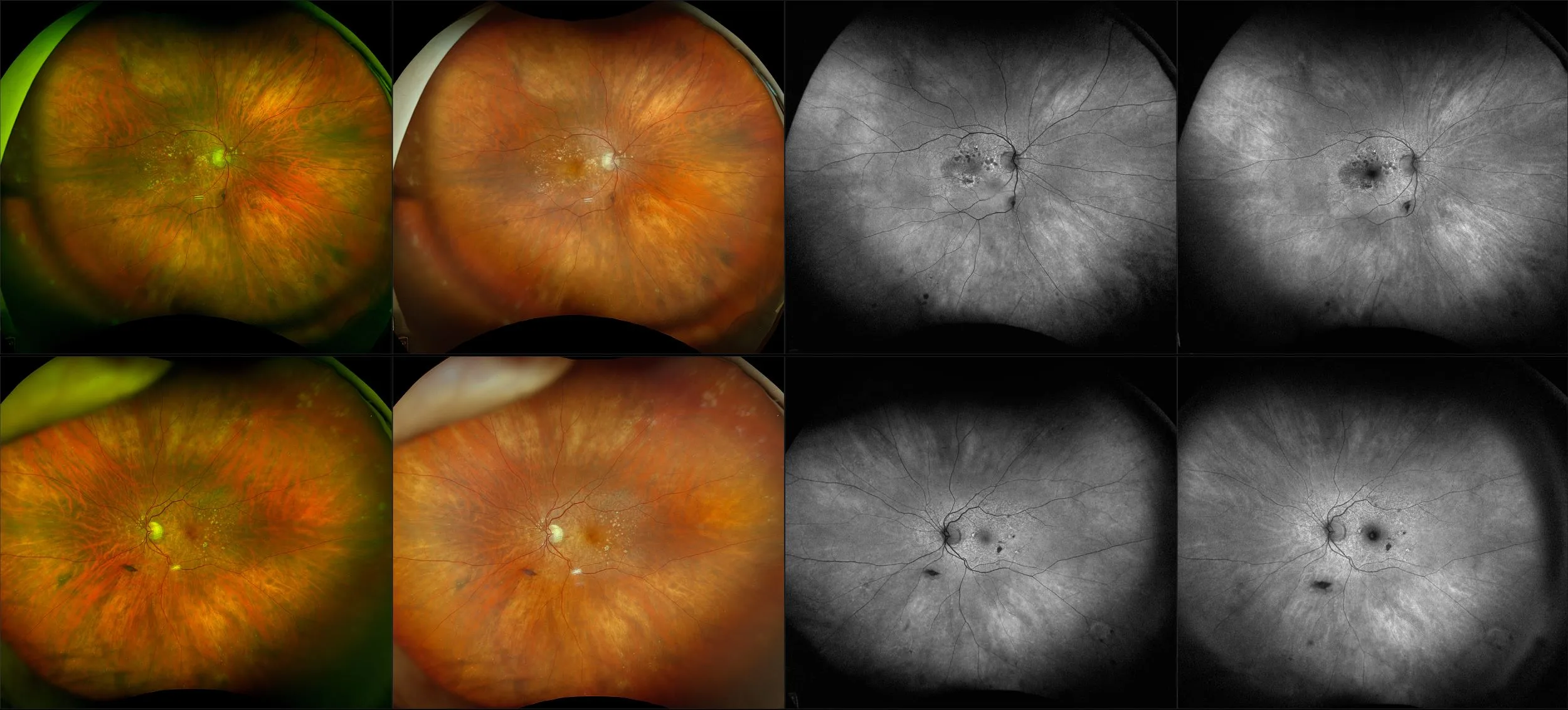
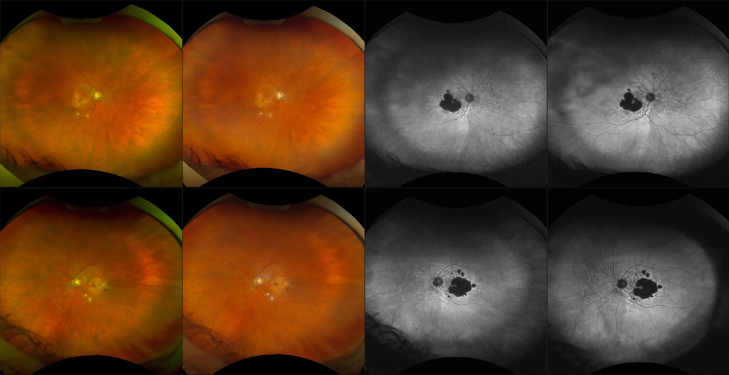
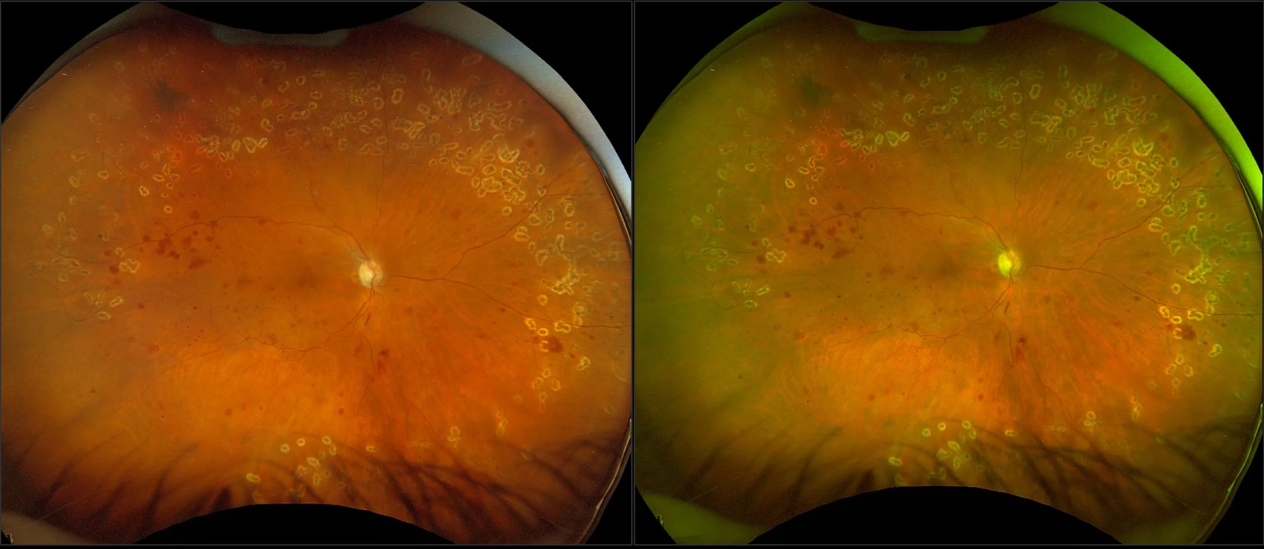
In a recent assessment of the new optomap color rgb vs. optomap color rg, Topcon white light color montage (dilated) and Heidelberg Multicolor (MCI) 55-degree steered images on 80 eyes with retinal, choroidal and optic nerve pathologies found that optomap color rgb was significantly superior in diagnostic information compared to gold standard Topcon and Heidelberg Multicolor.
“The results of our study show that the new Optos California provides a well-balanced color image, while users can choose to use both the color rg or color rgb images to enhance visualization of vitreous, retinal, and choroidal structures.” - OSLI, 2023
We encourage you to contact us if you have questions regarding how optomap and Optos technology can benefit your patients and clinical settings.
Clinical Studies
Comparison of a Novel Ultra-Widefield Three-Color Scanning Laser Ophthalmoscope to Other Retinal Imaging Modalities in Chorioretinal Lesion Imaging.
New 200° Single-Capture Color Red-Green-Blue Ultra-Widefield Retinal Imaging Technology: First Clinical Experience.
This new device allows for both RG and RGB imaging of the retina, providing valuable information on the anatomy of the vitreoretinal interface, retina, retinal pigment epithelium, and their abnormalities. Both techniques complement each other and can be useful in daily practice, allowing clinicians to choose the preferred imaging technique depending on specific findings and conditions.
Optos unveils ultra-widefield color image modality, offering increased retinal visualization to ophthalmologists.
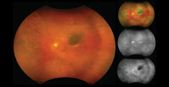
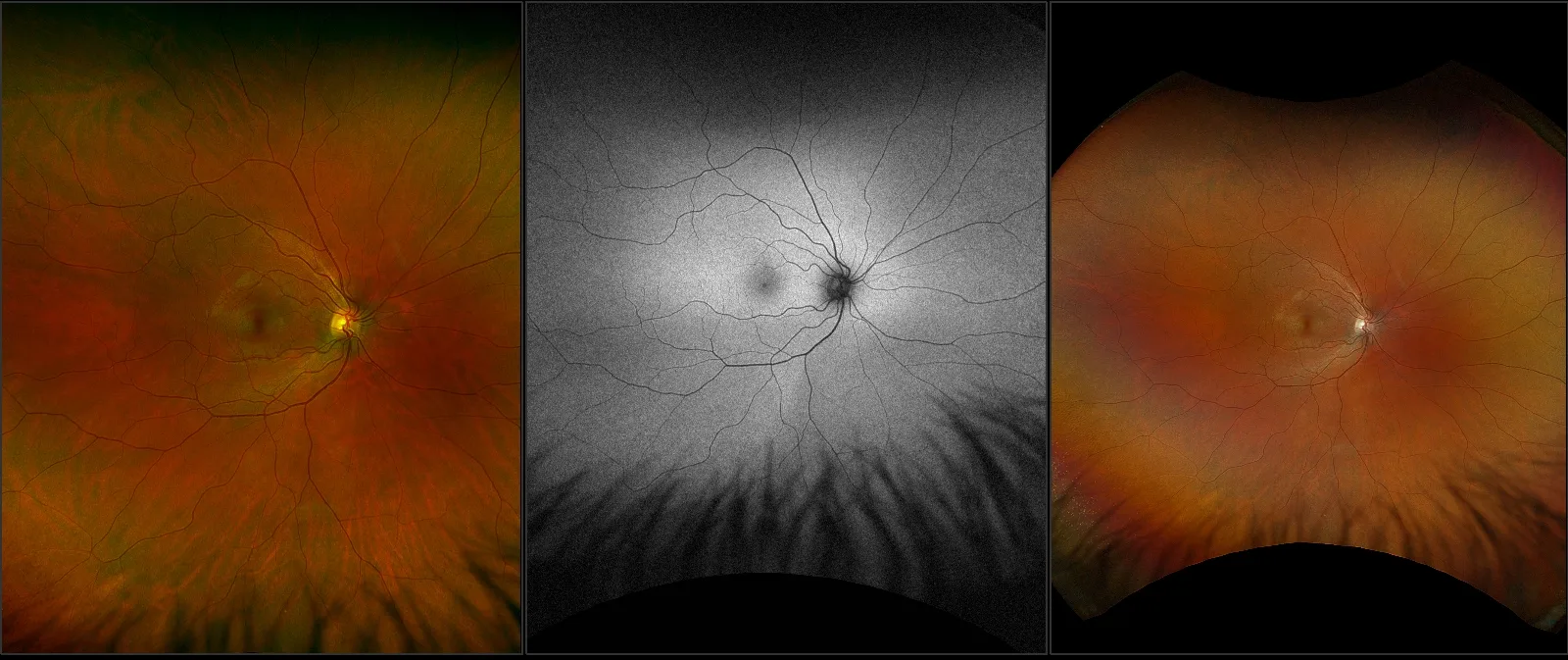
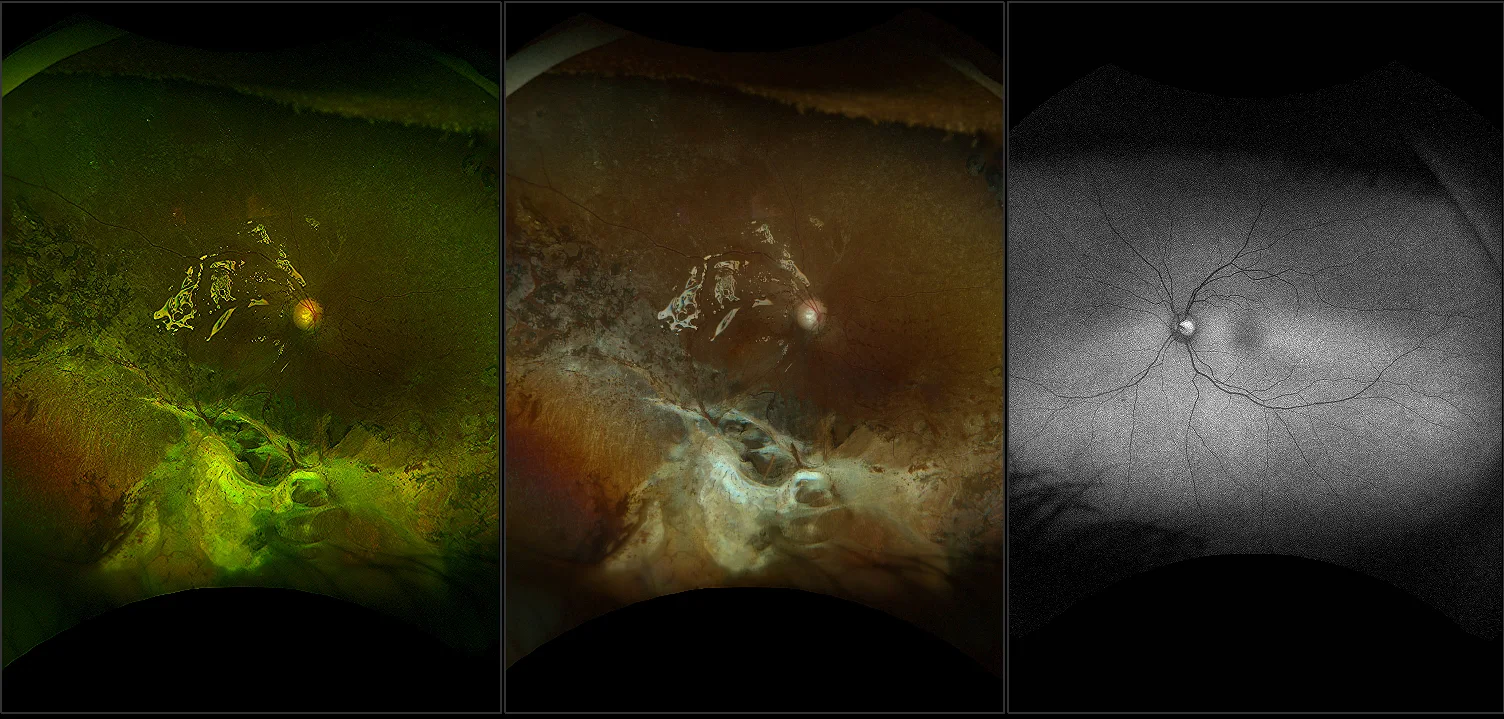
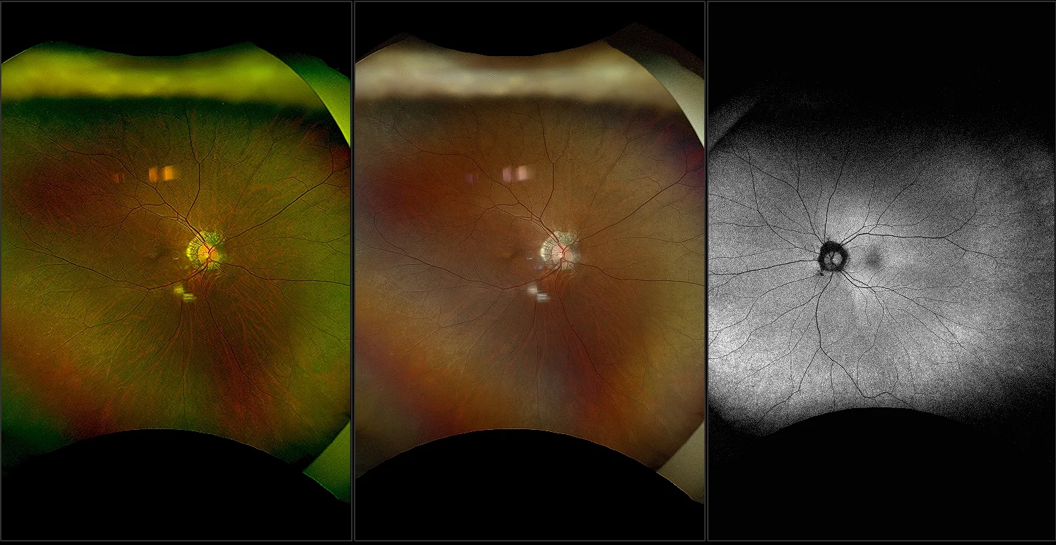
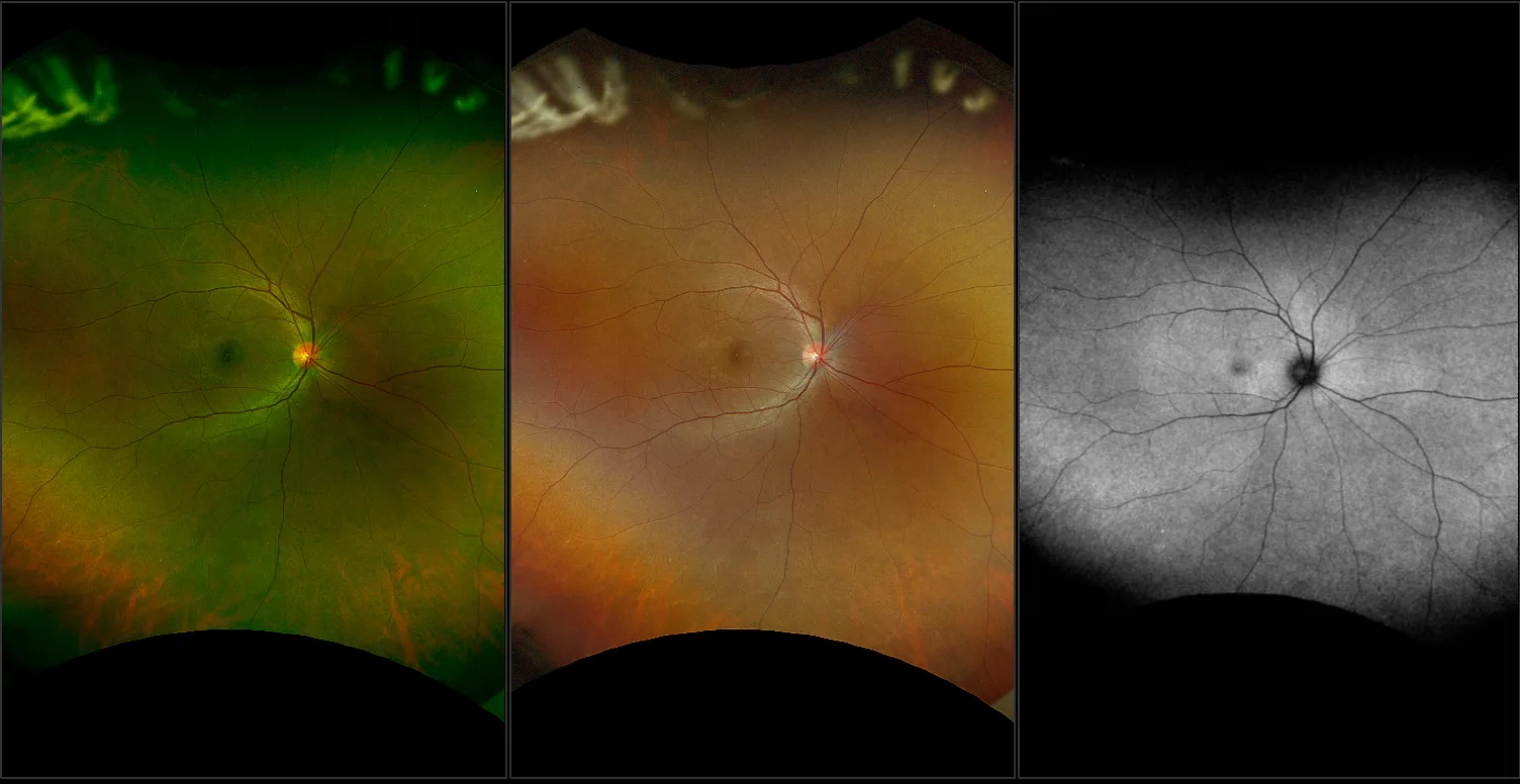
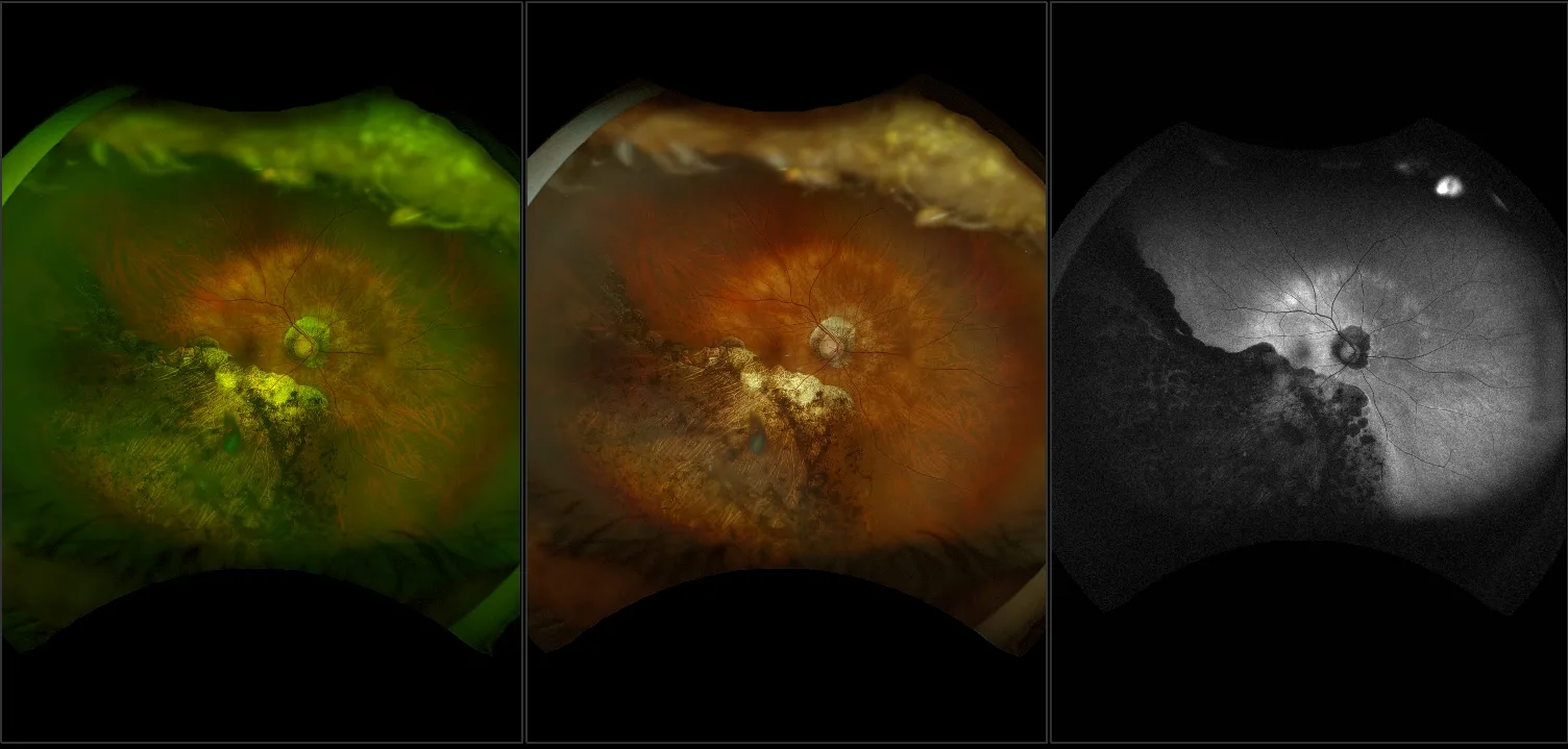
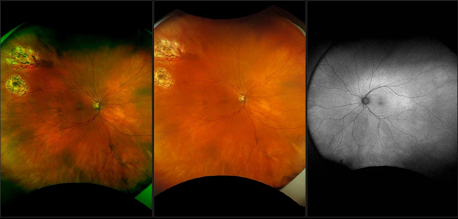
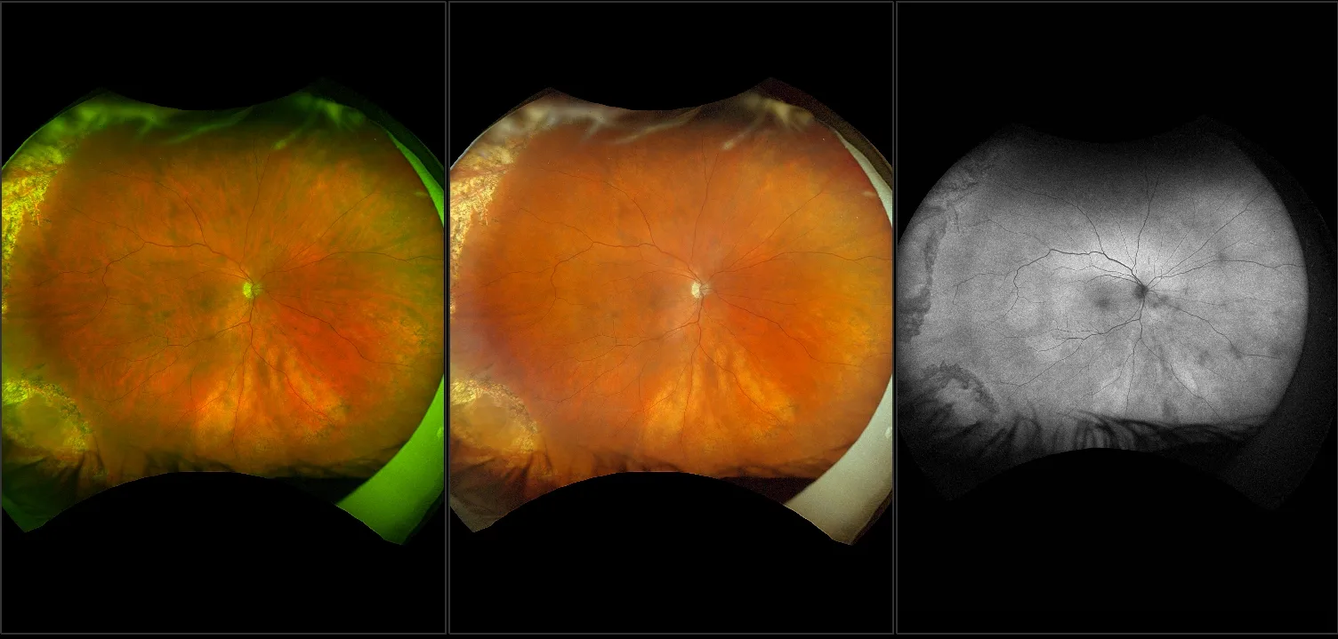
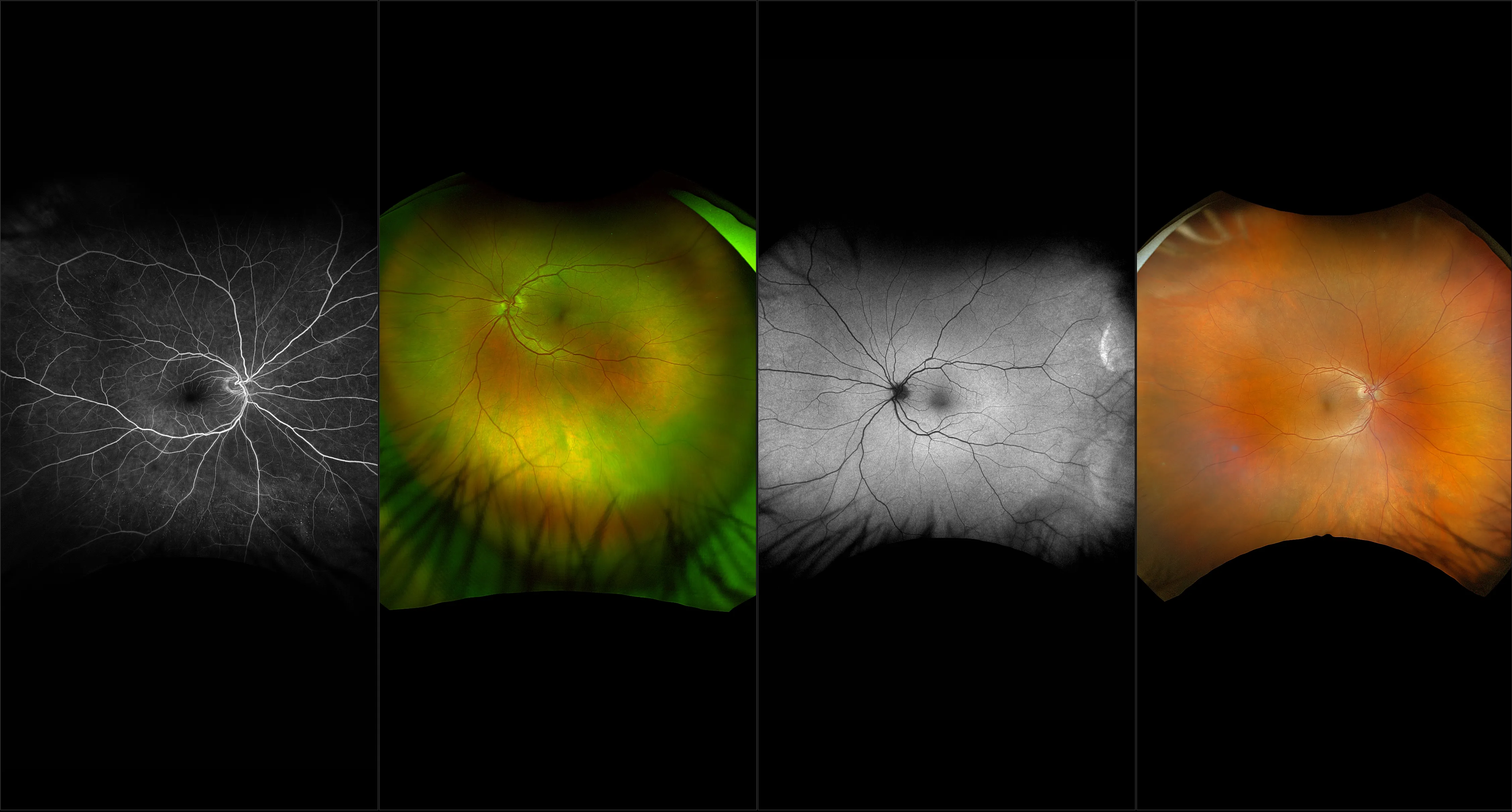
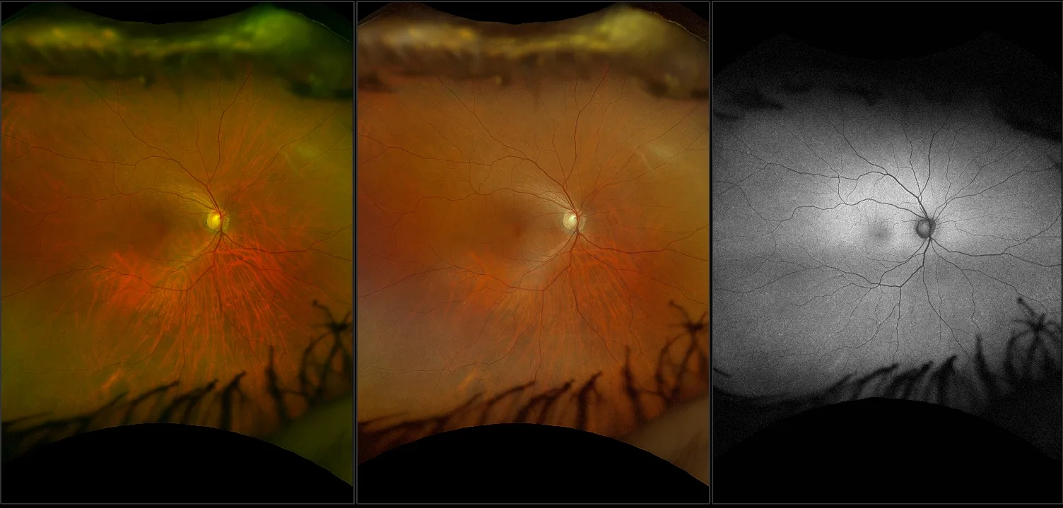
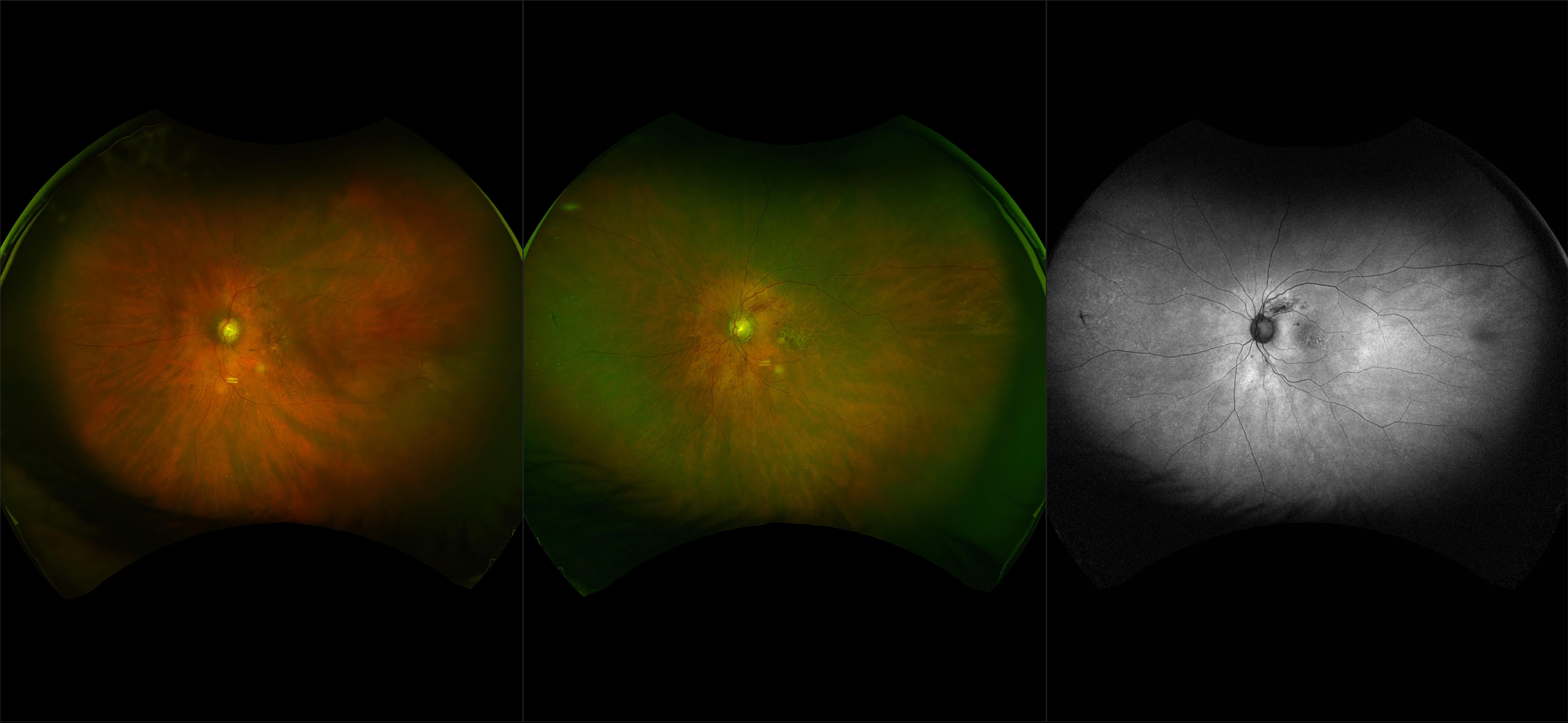
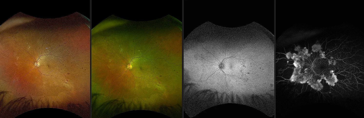
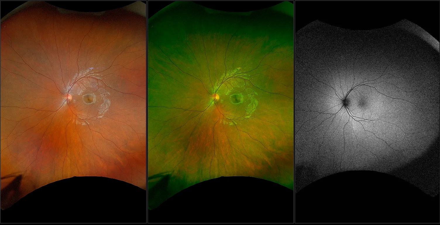
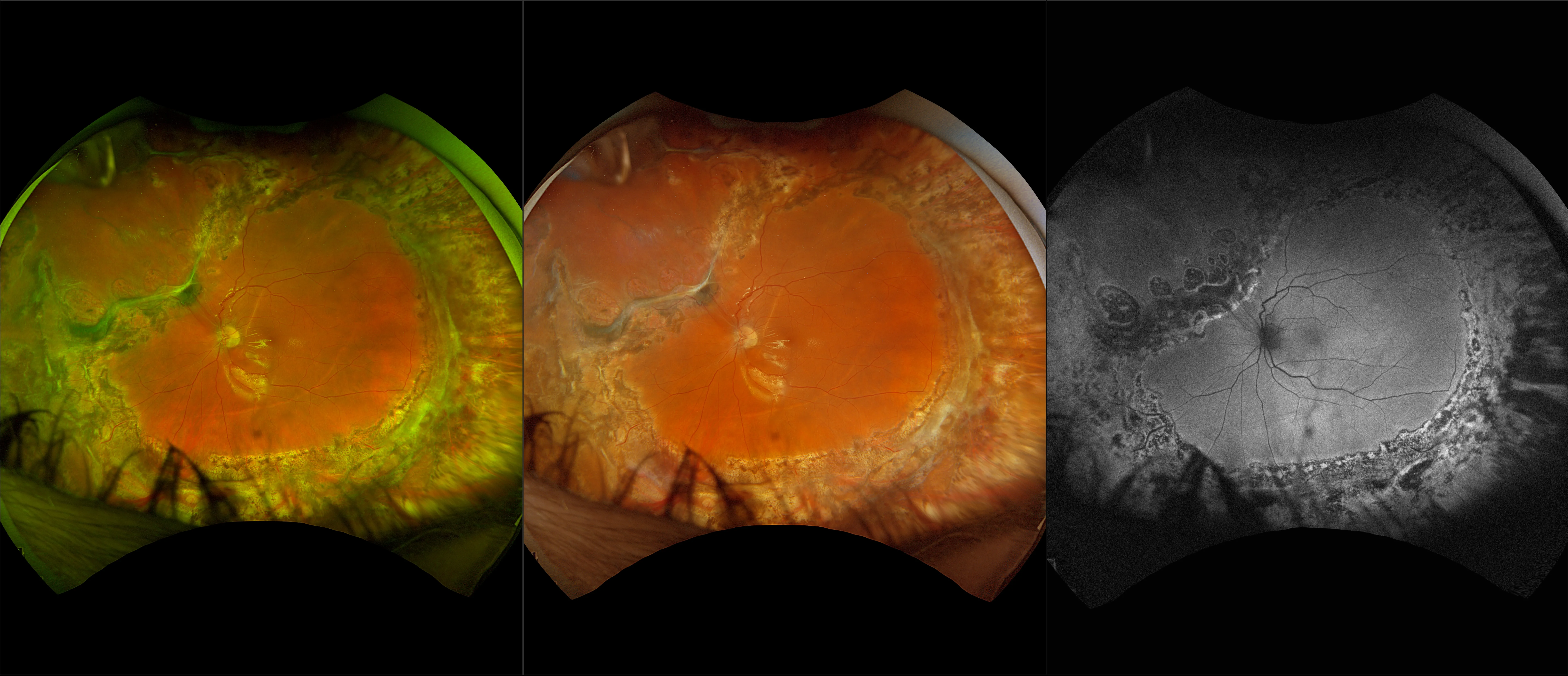
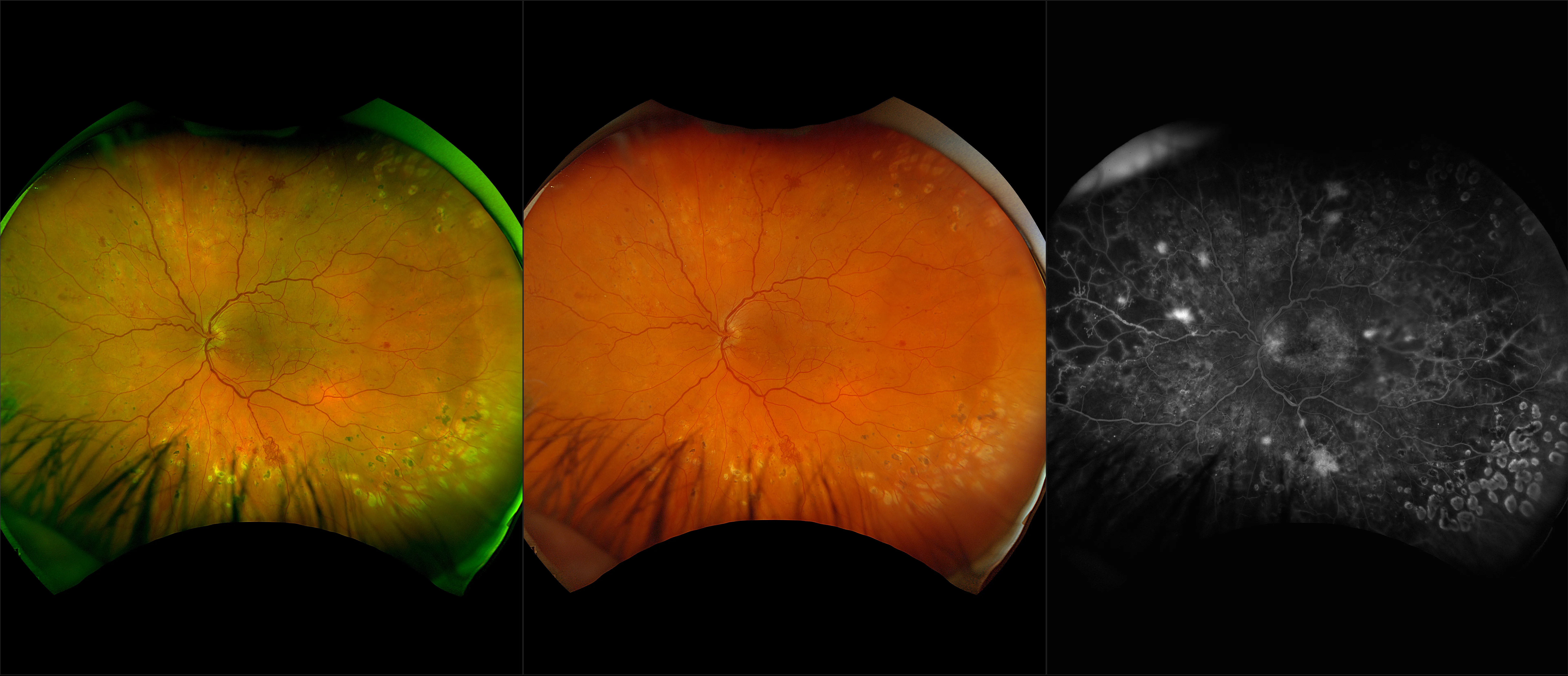
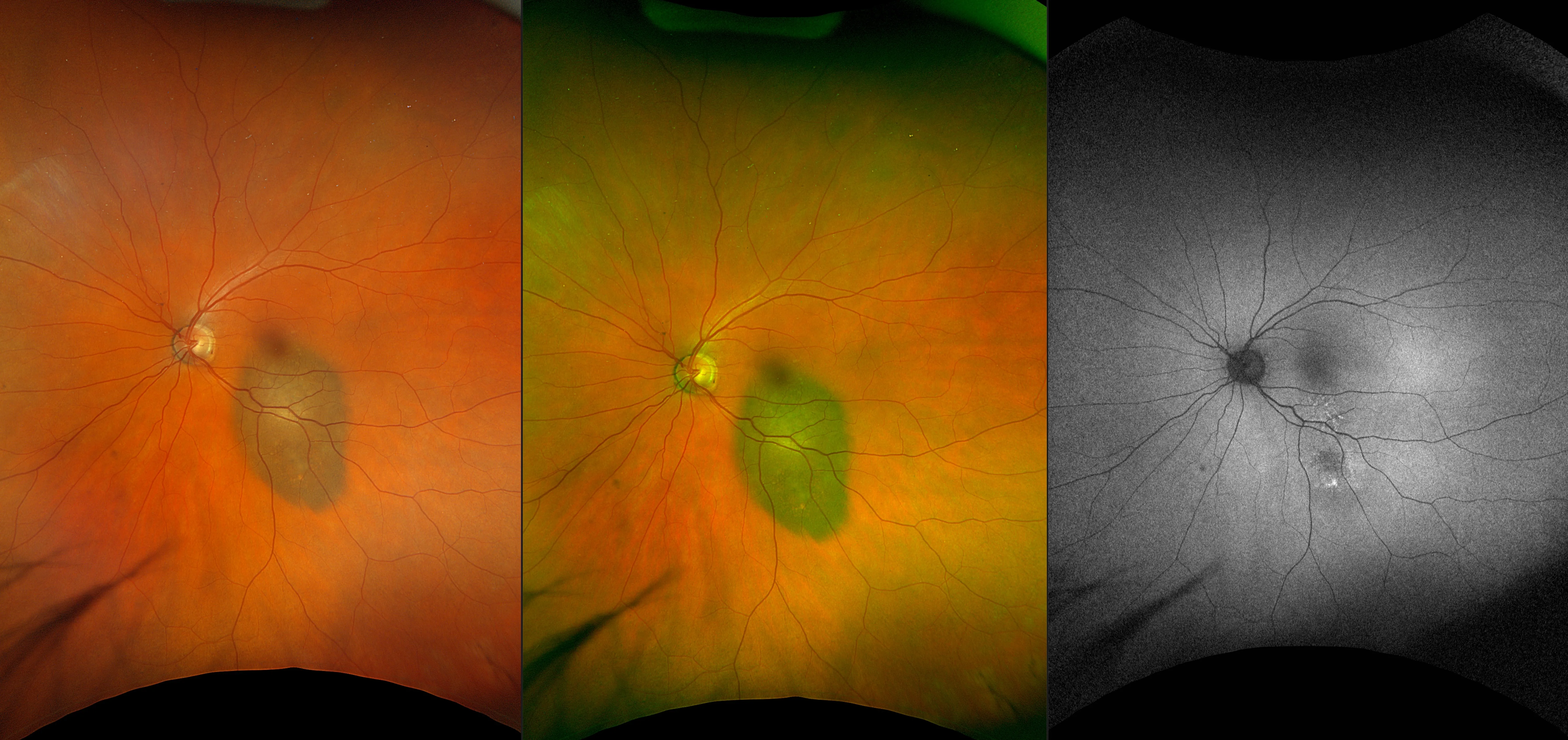
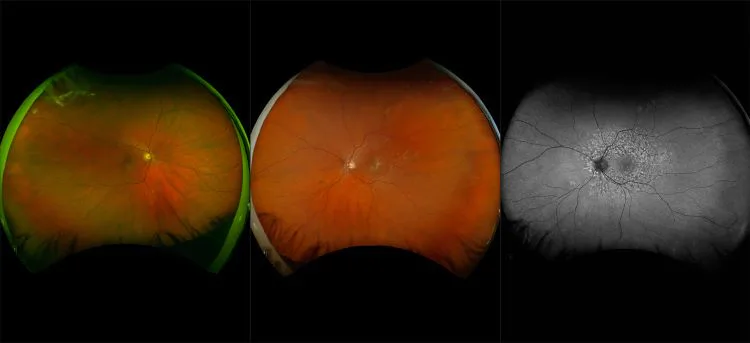
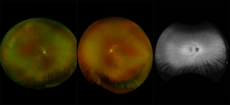
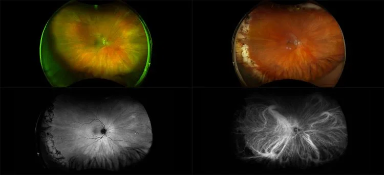
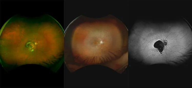
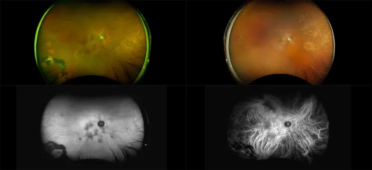
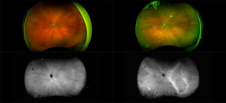
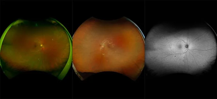
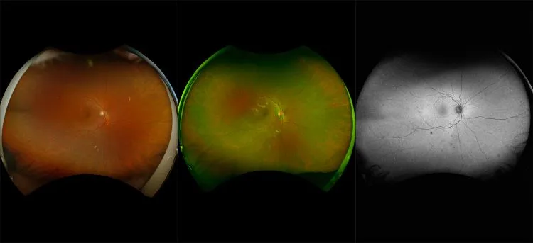
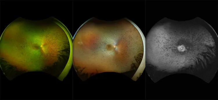
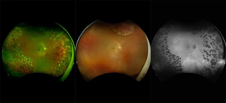
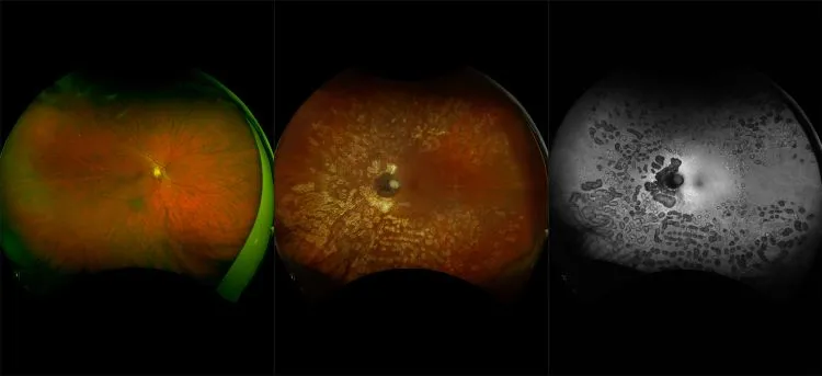
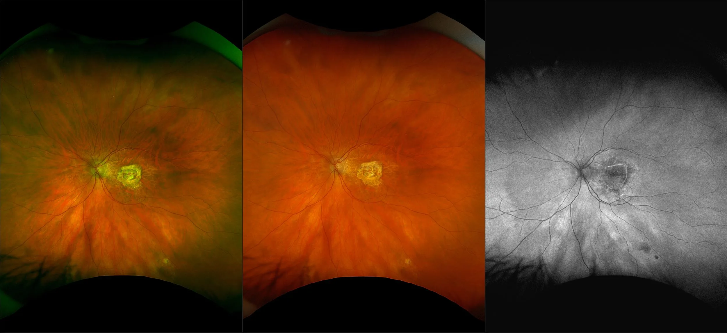
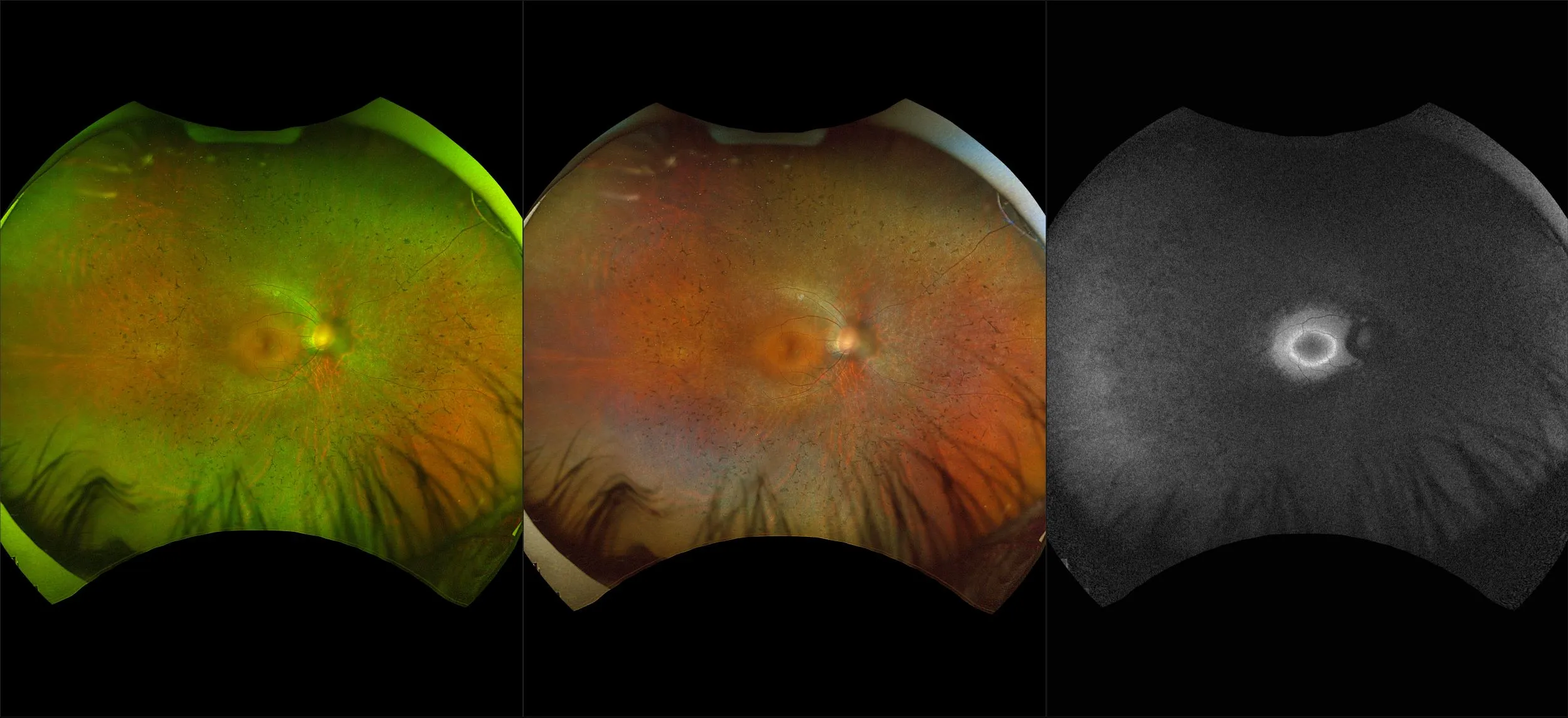
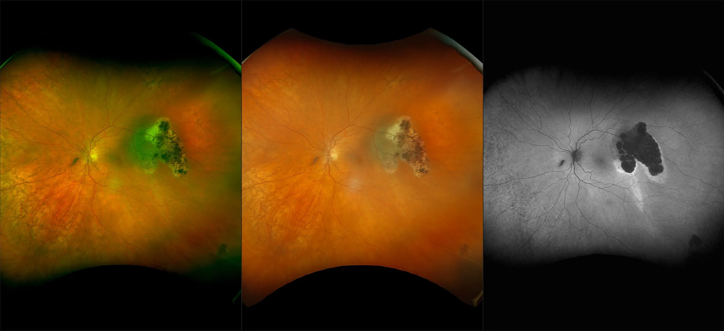
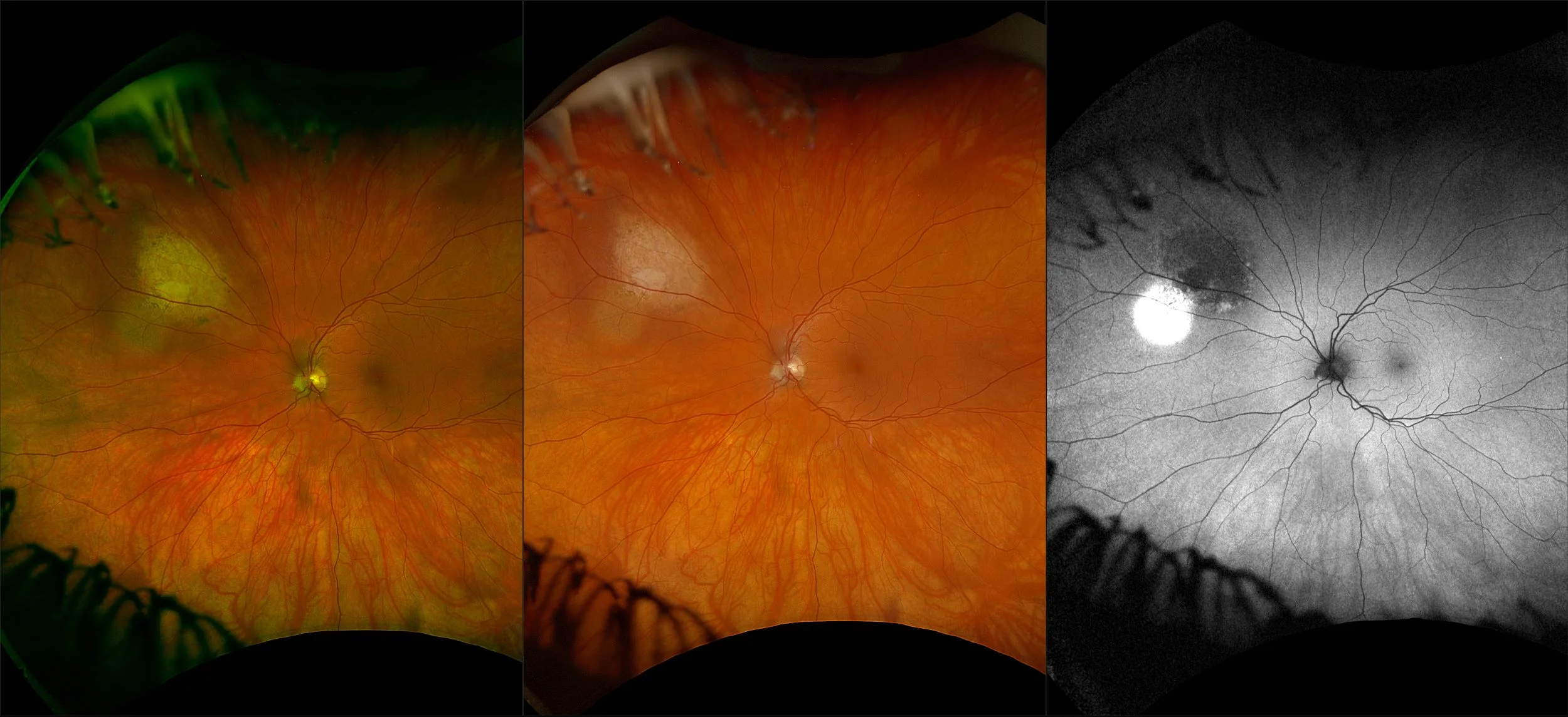
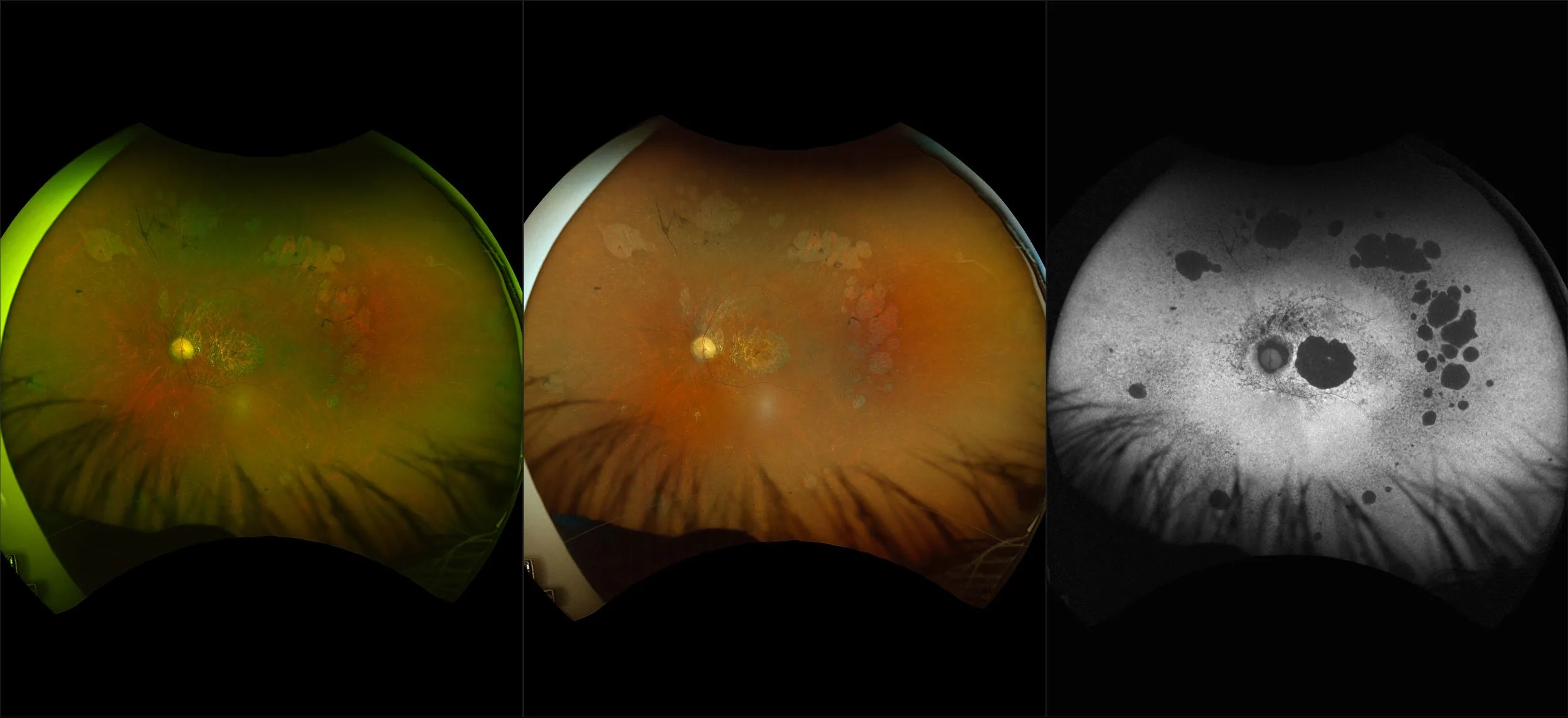
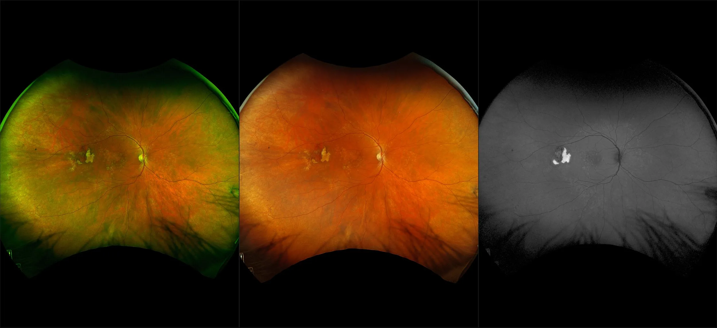
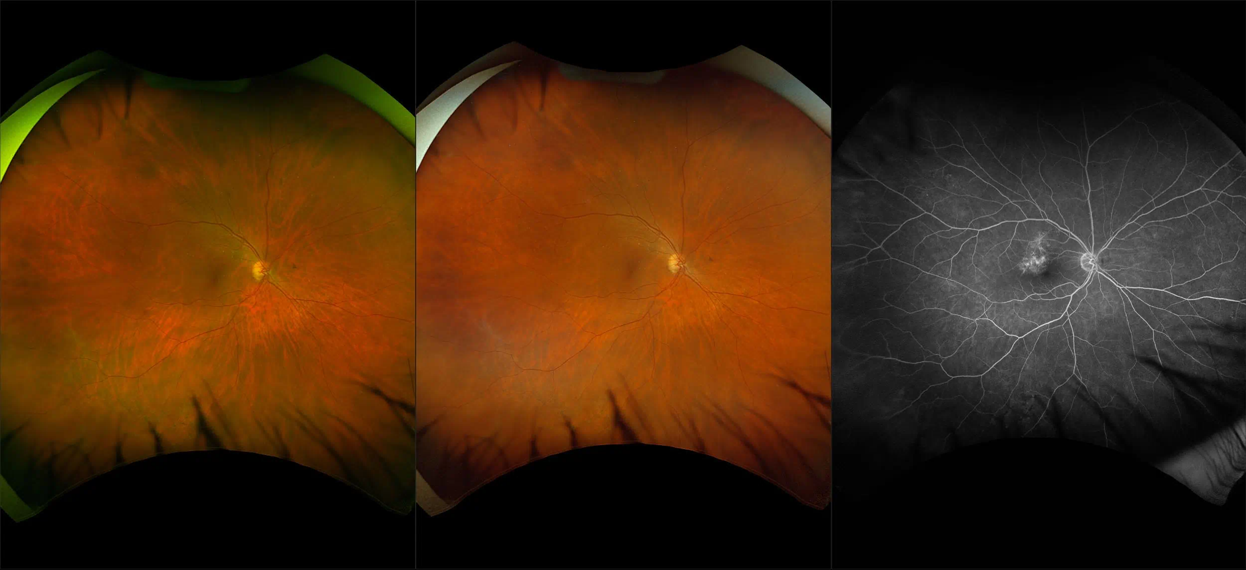
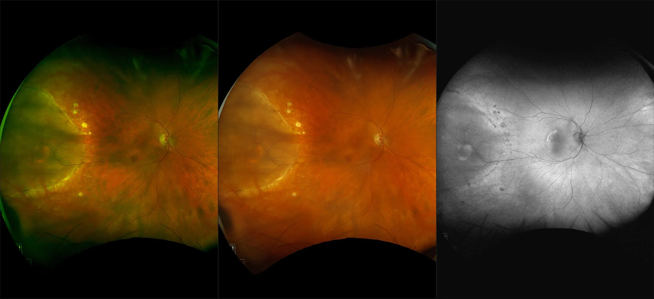
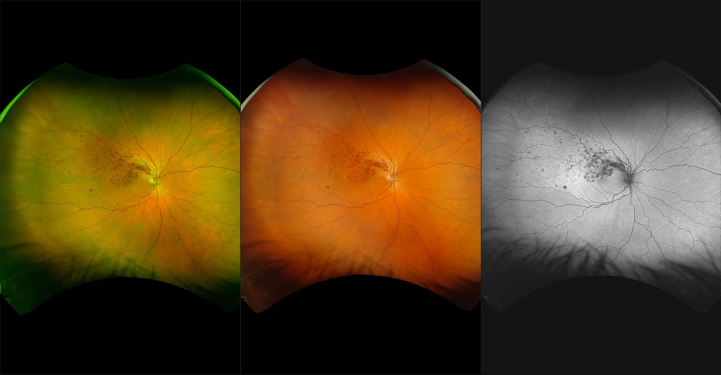
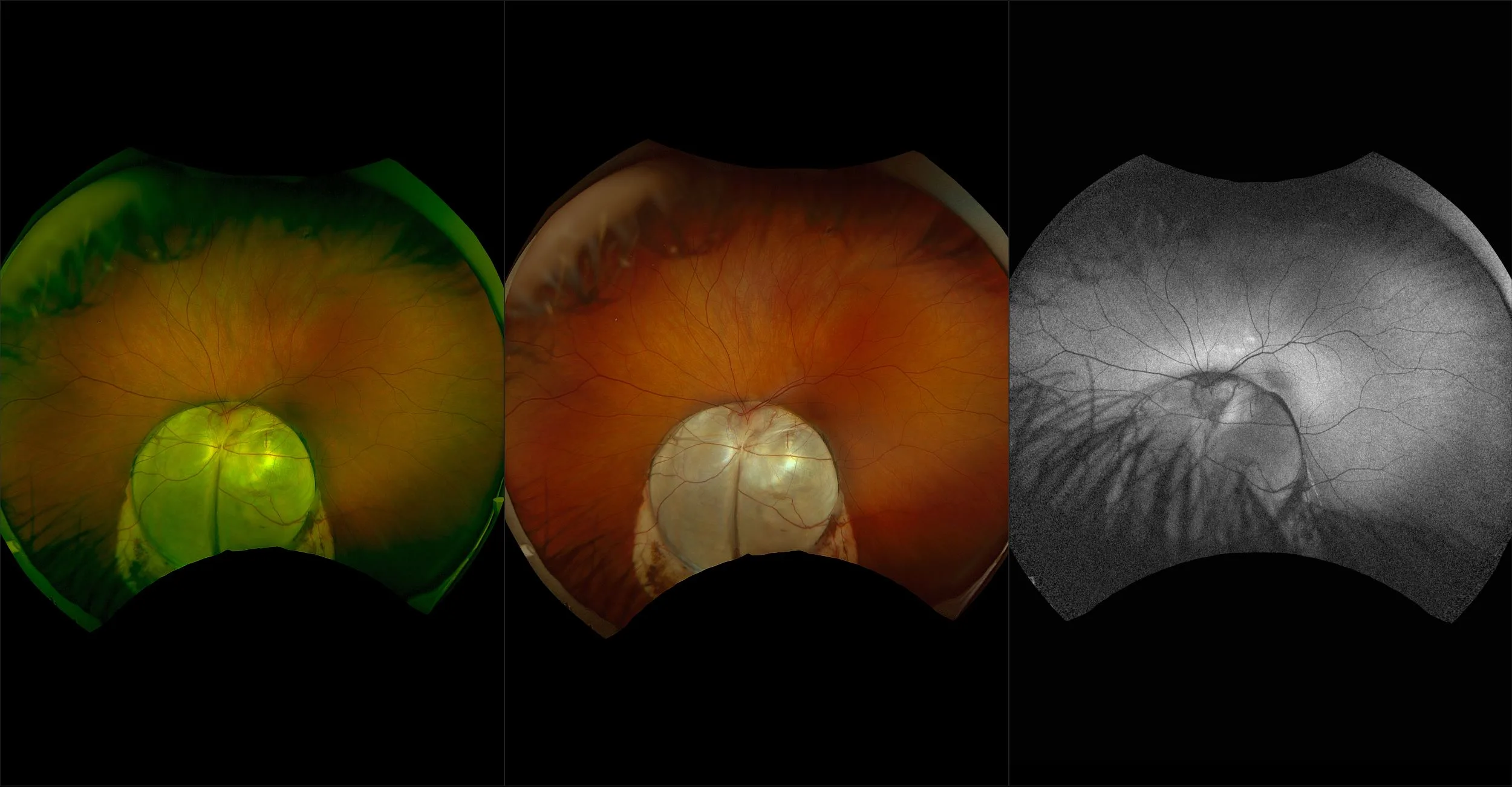
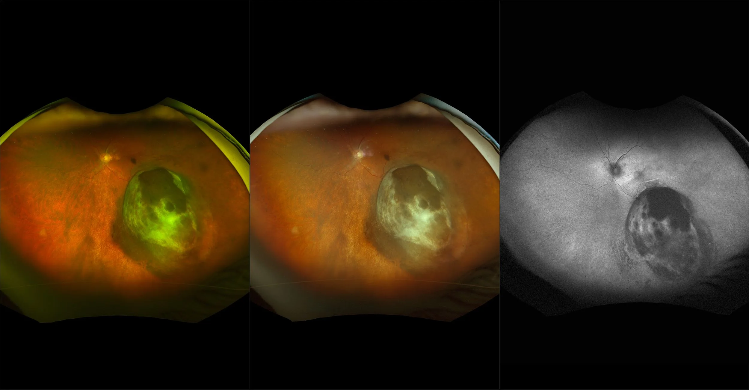
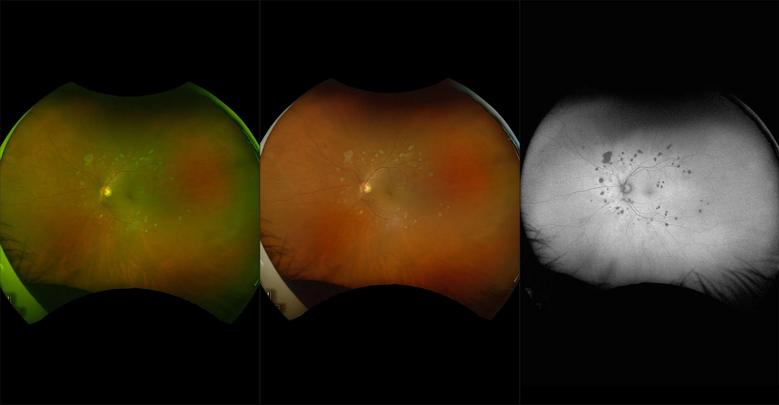
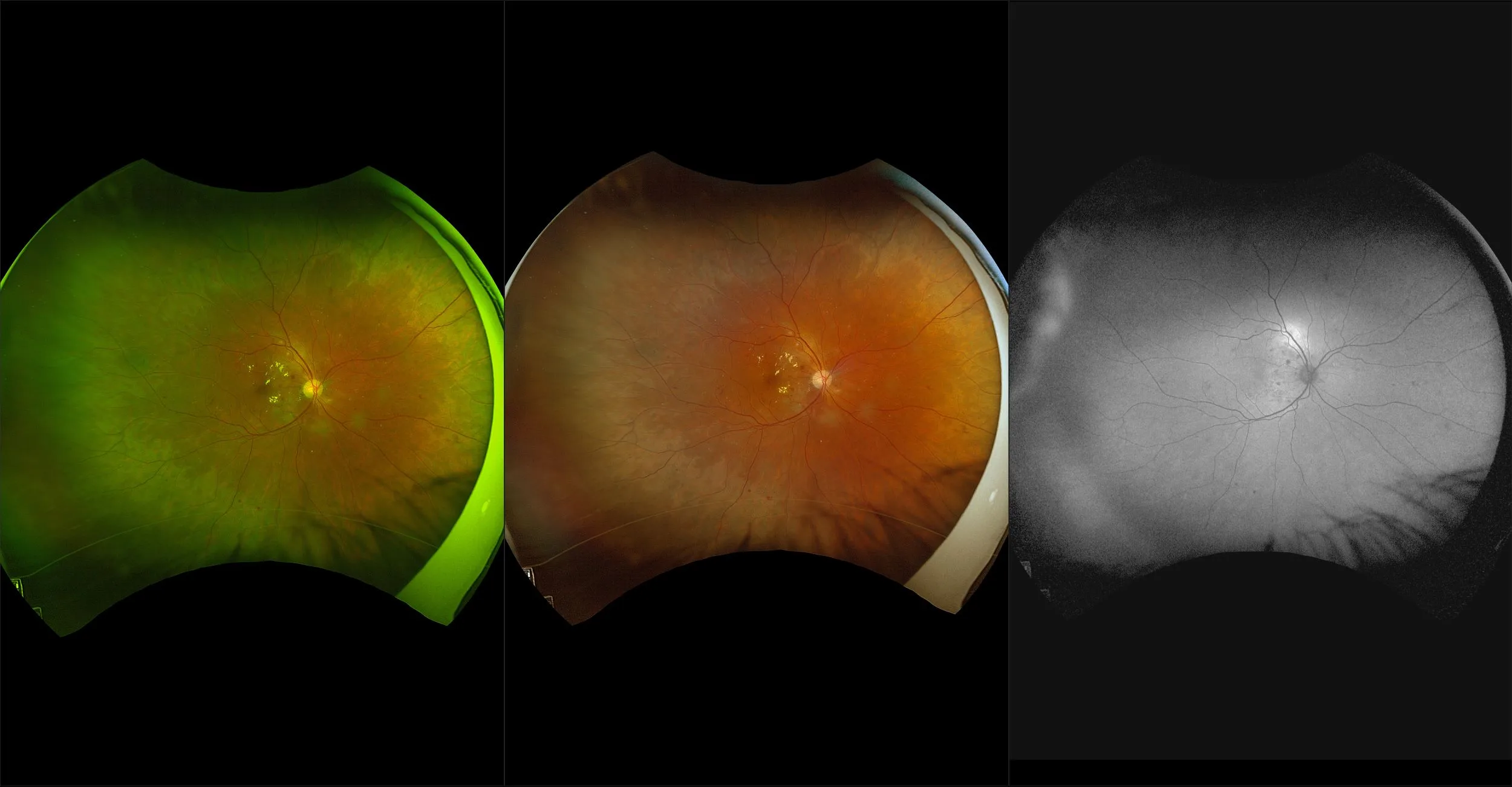
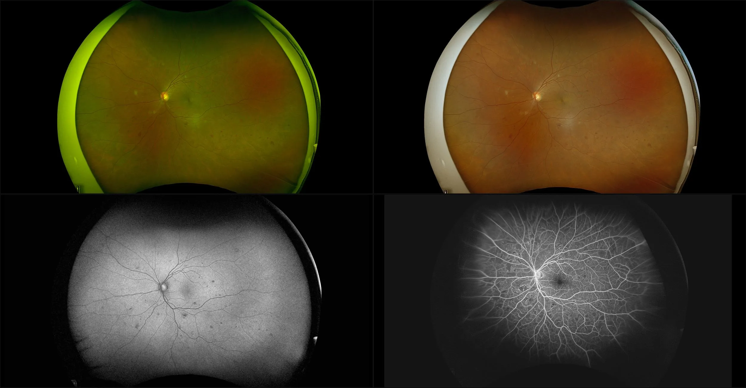
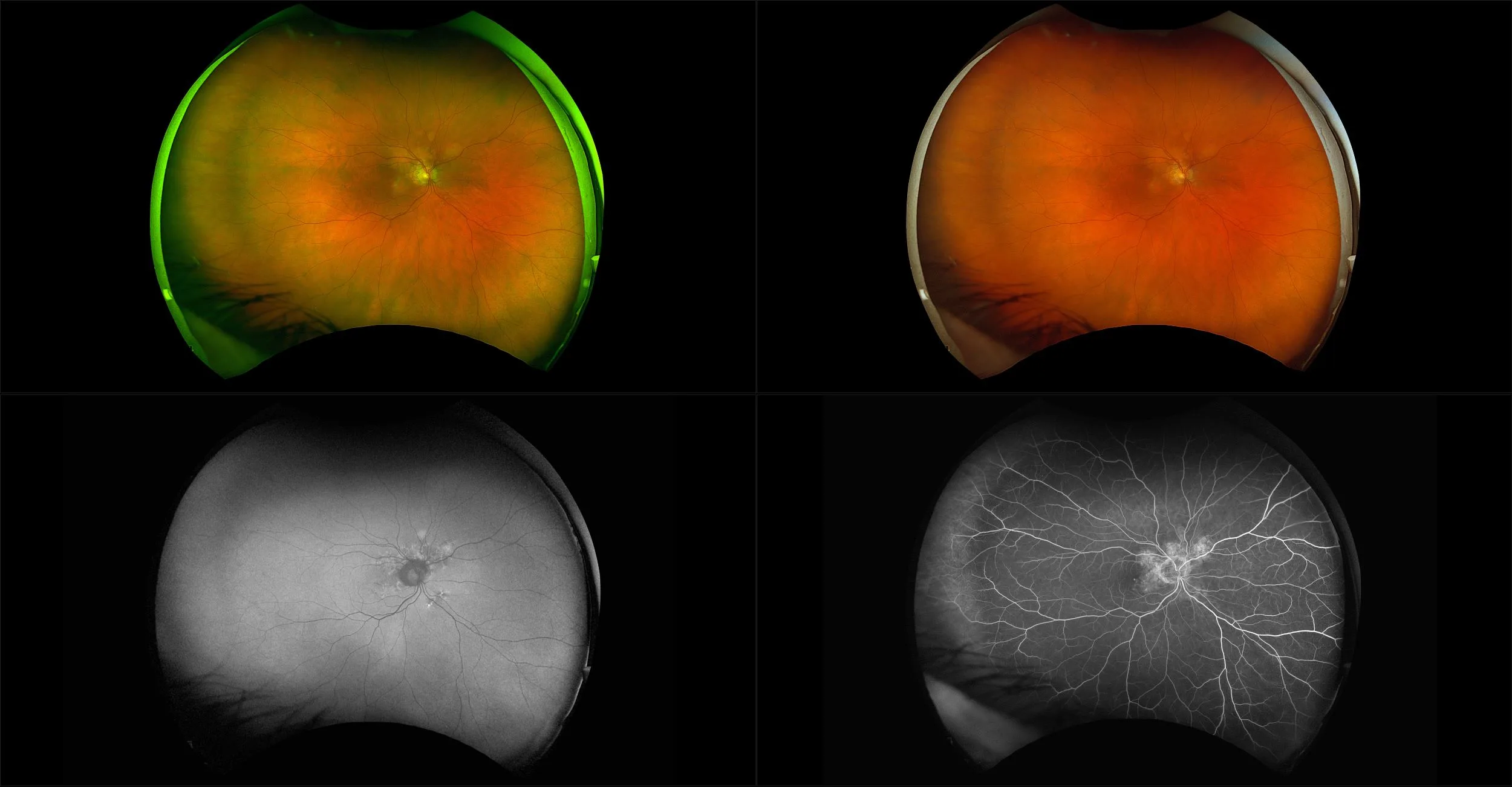
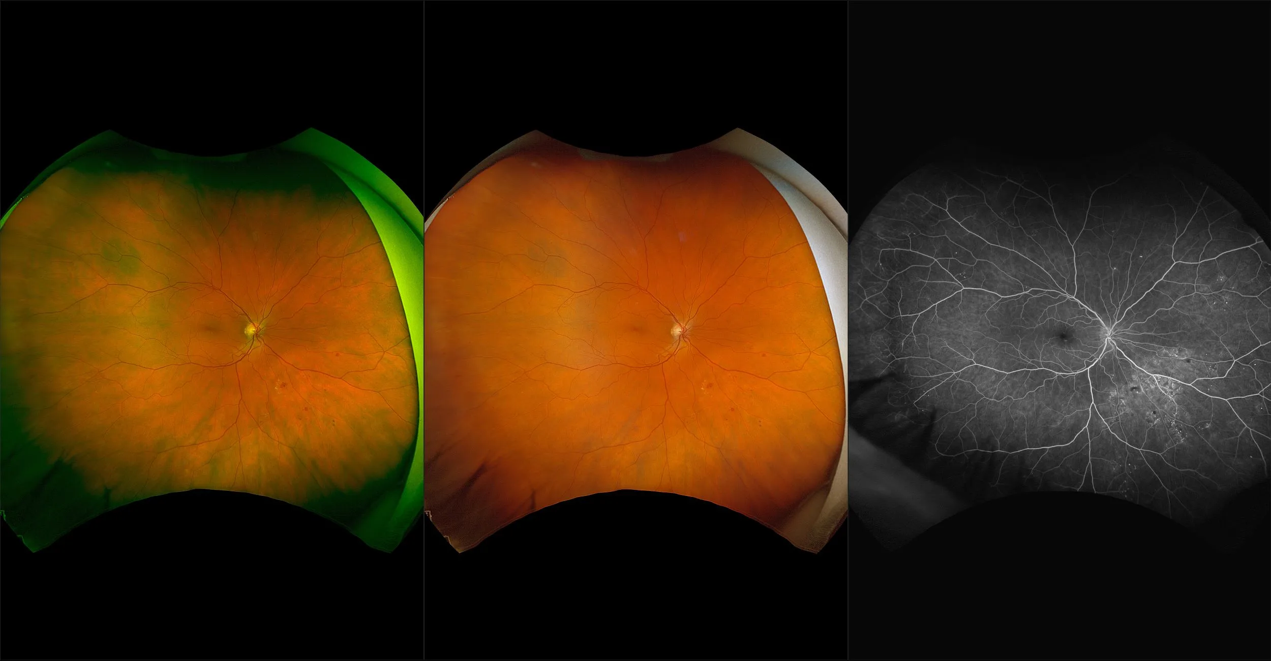
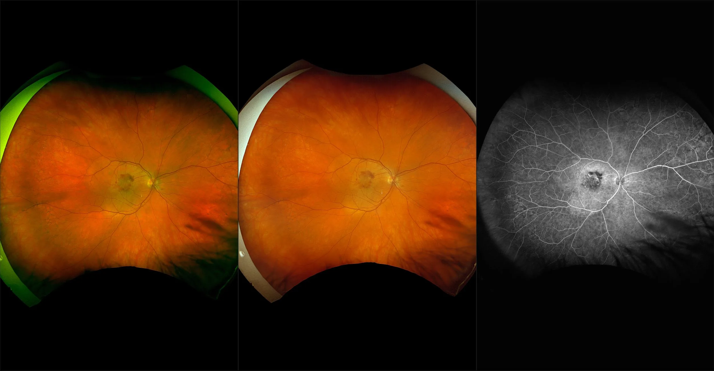
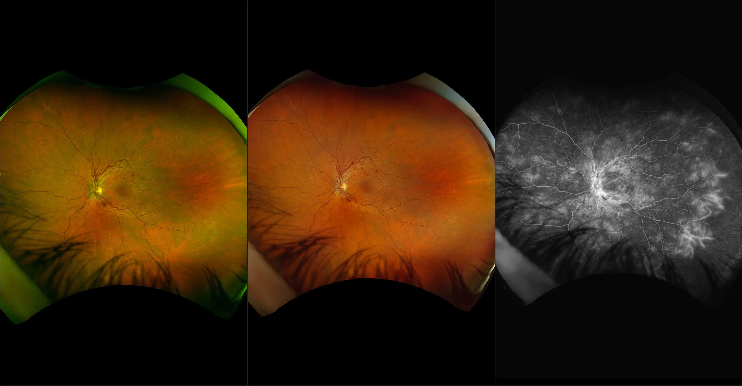
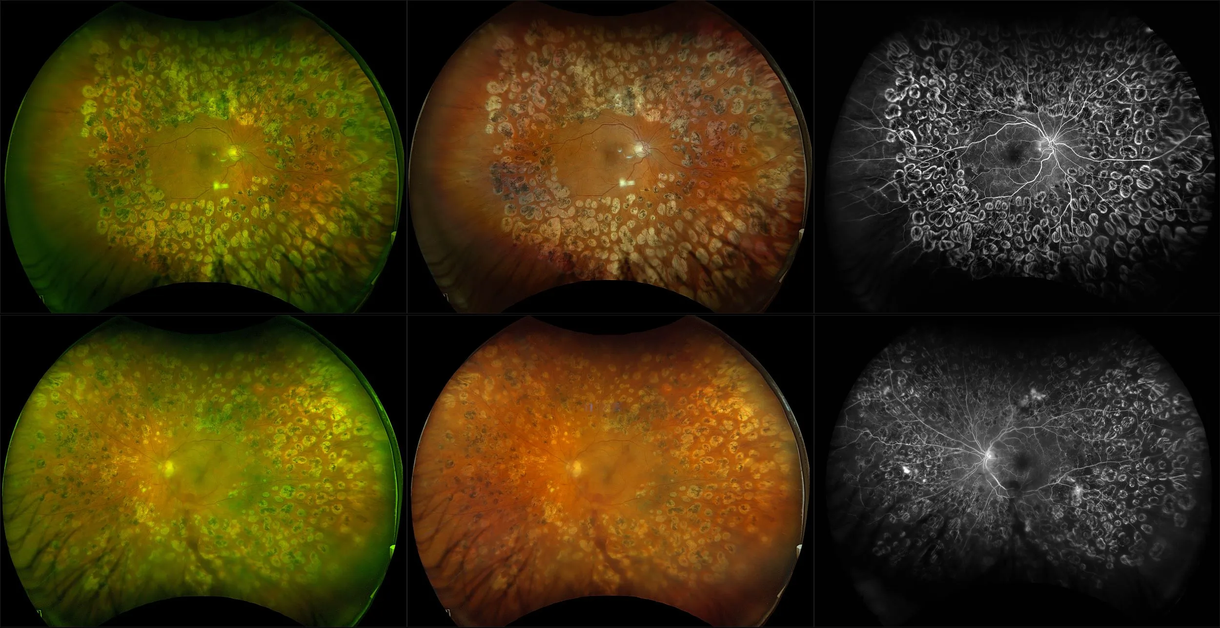
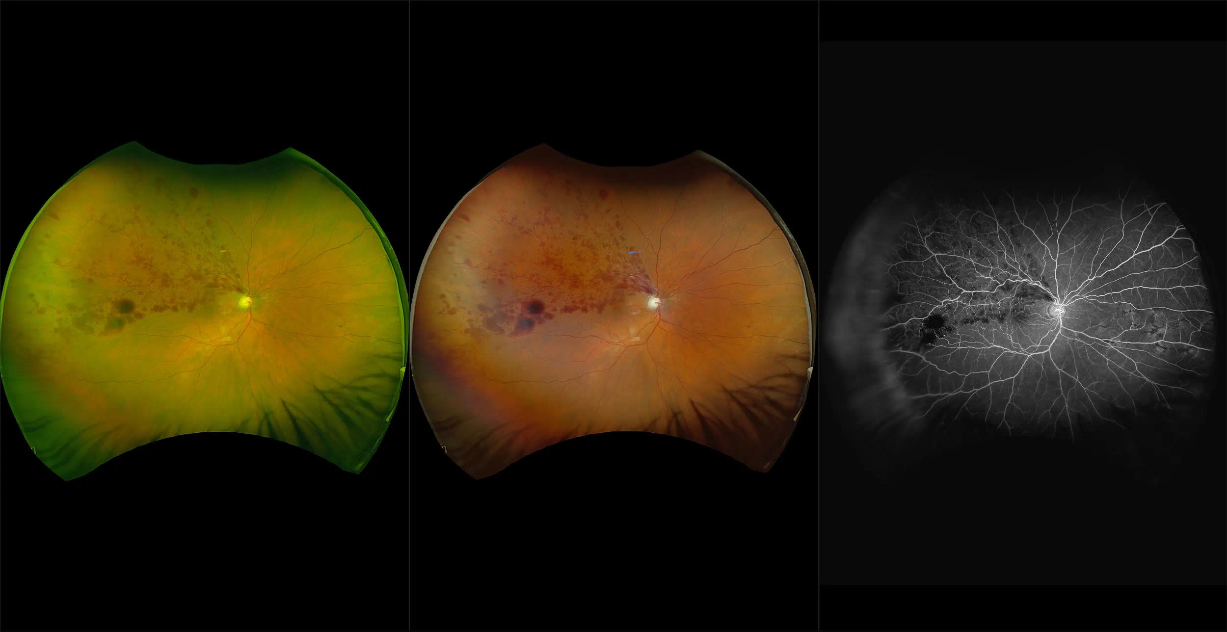
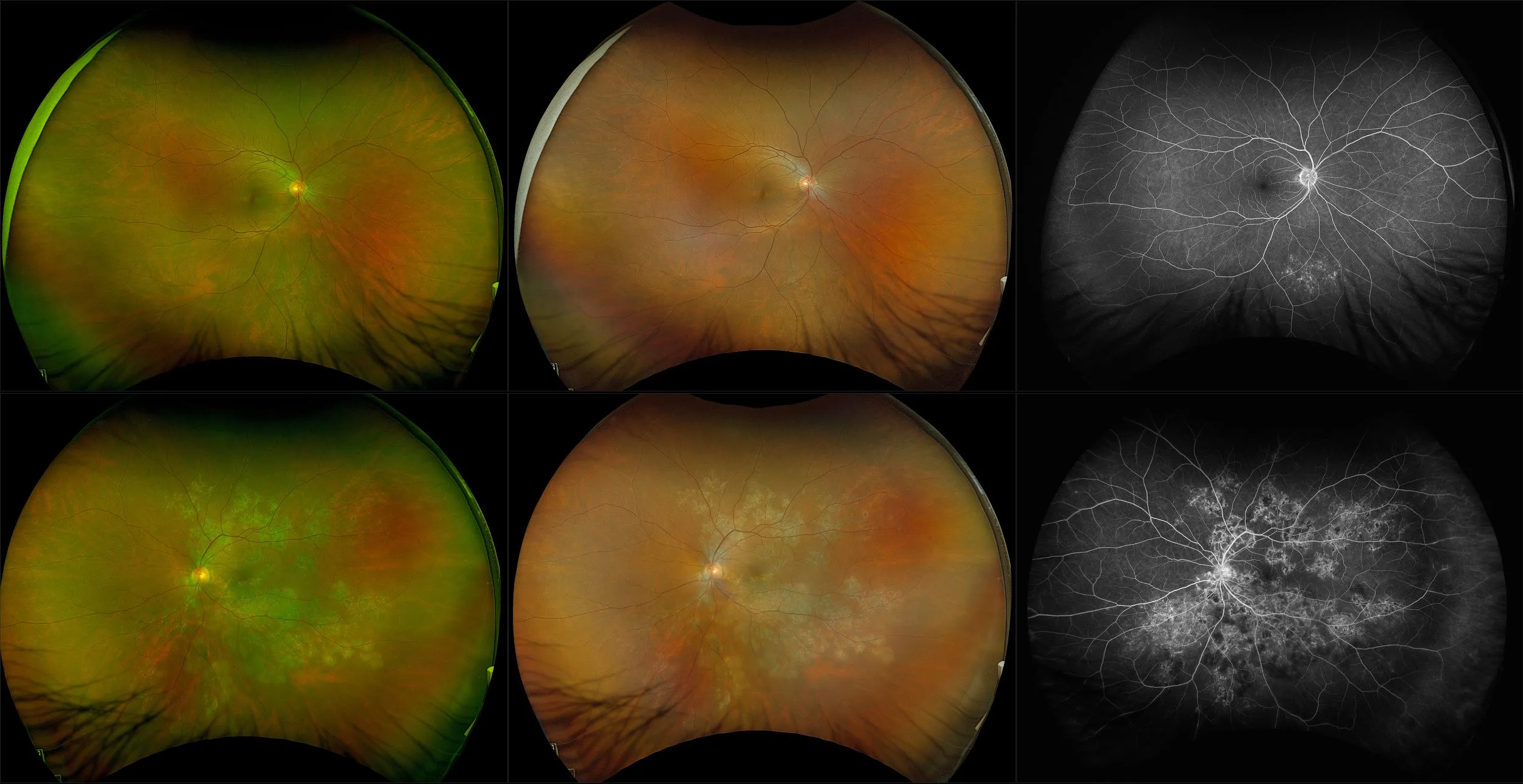
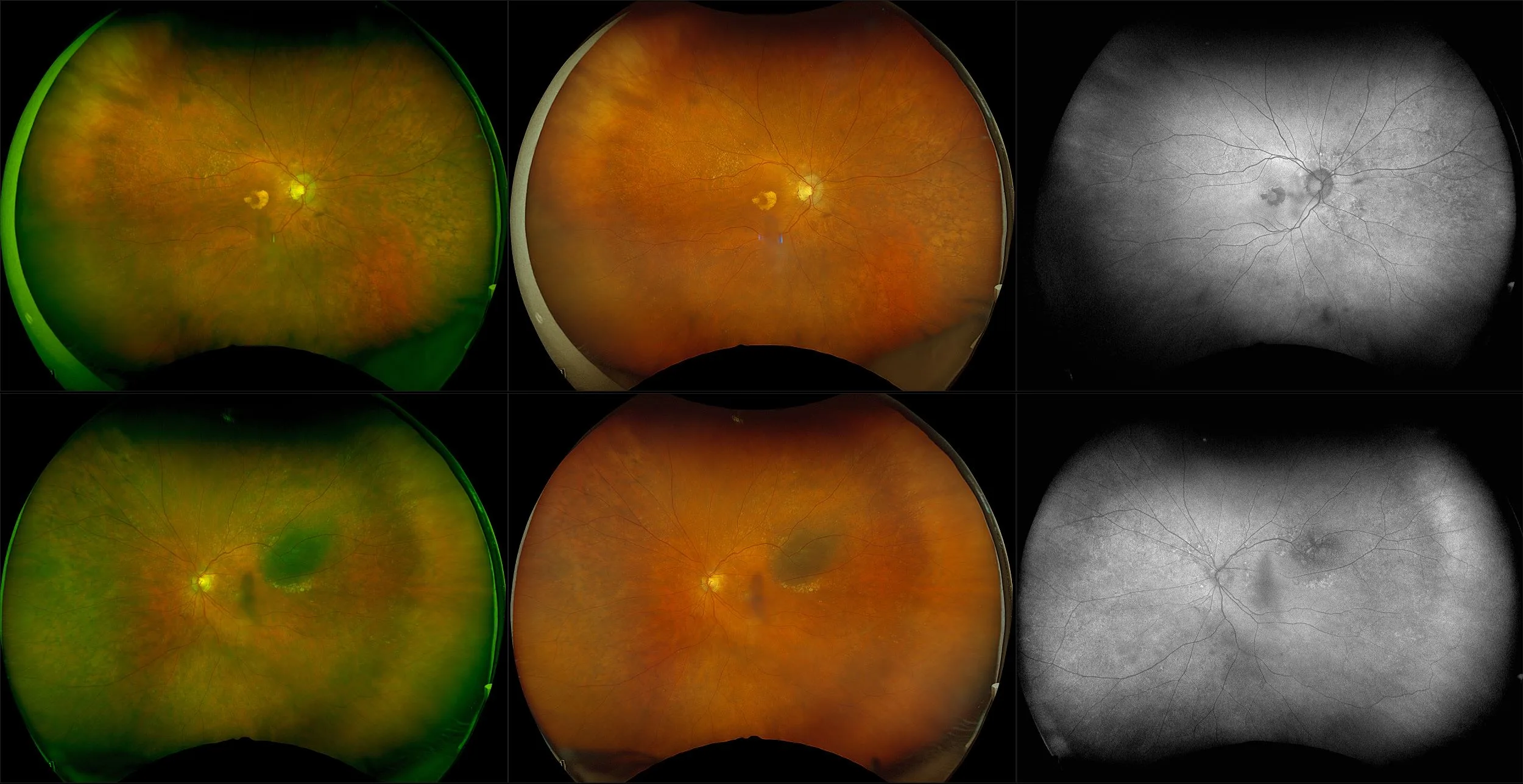
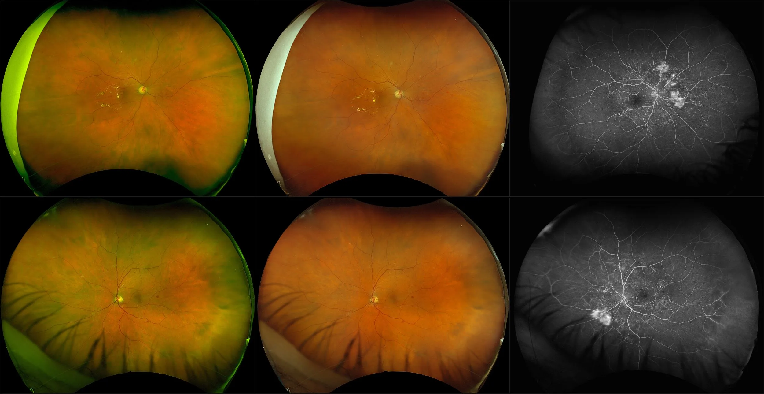
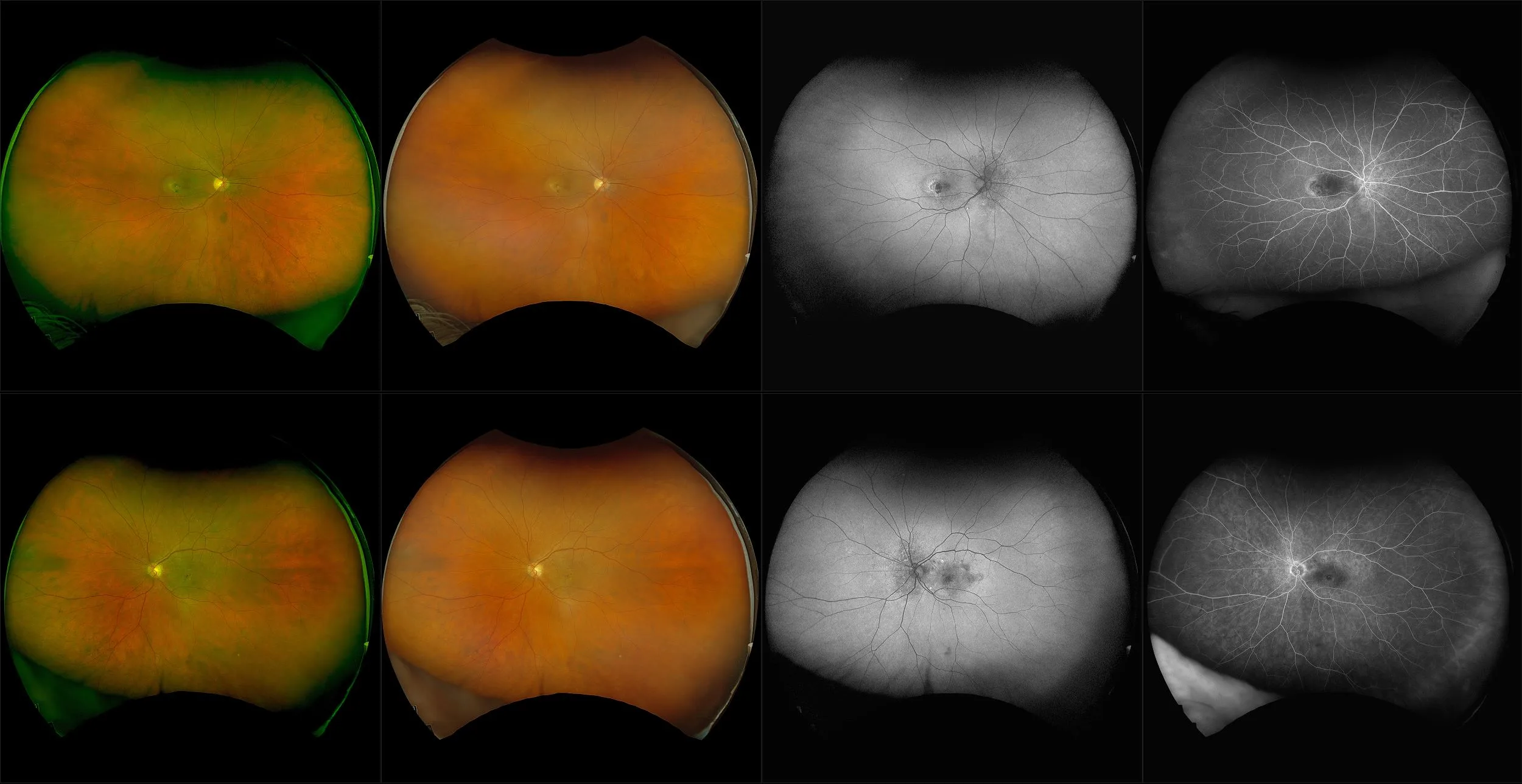
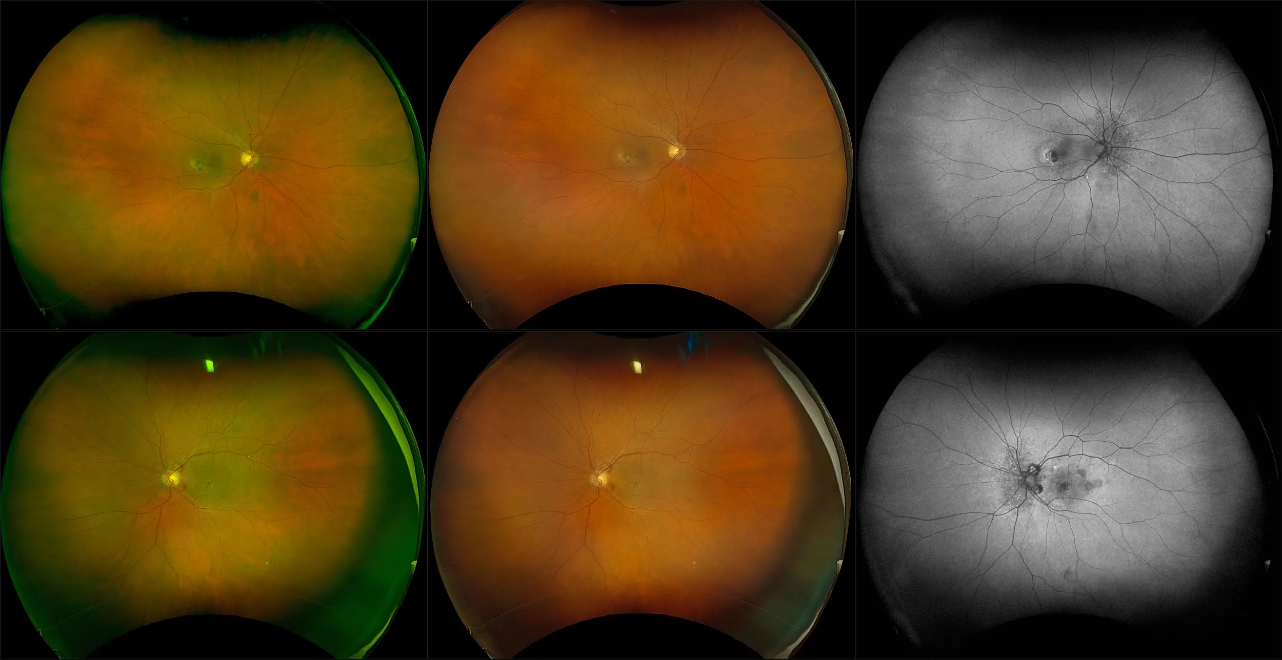
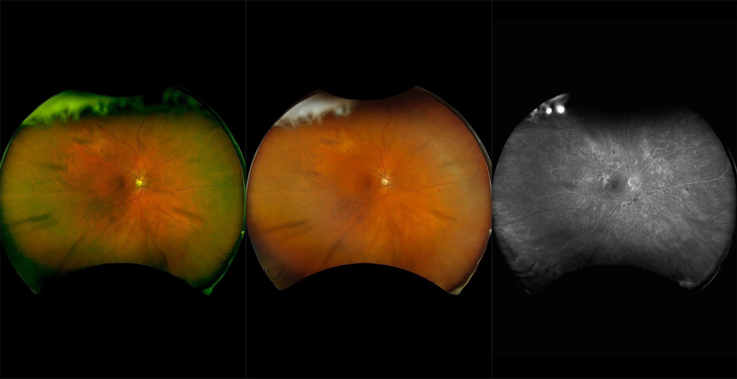
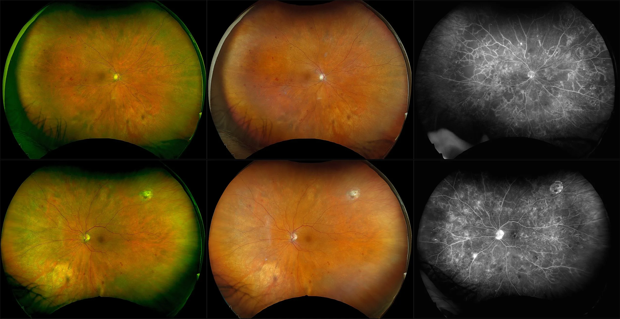
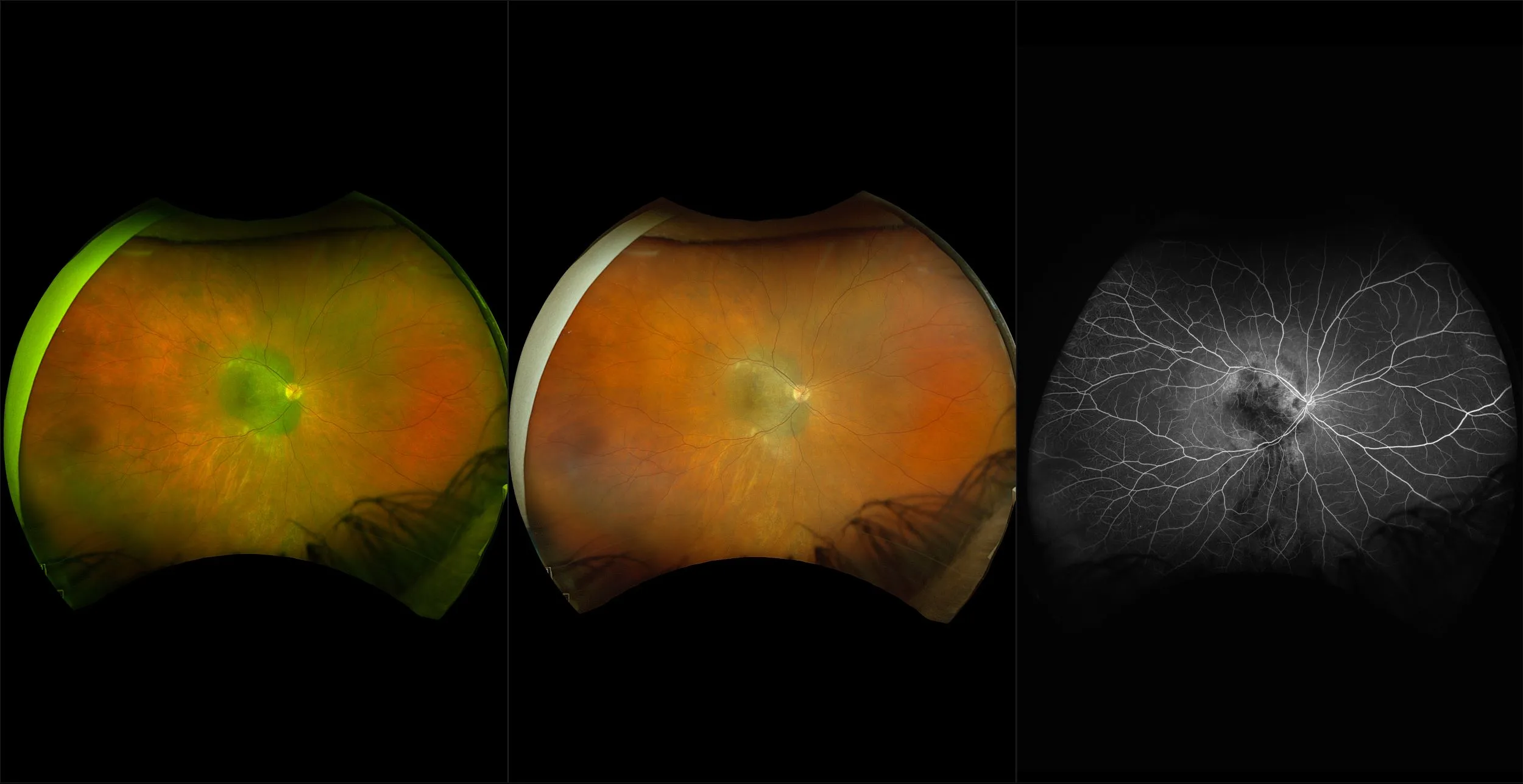
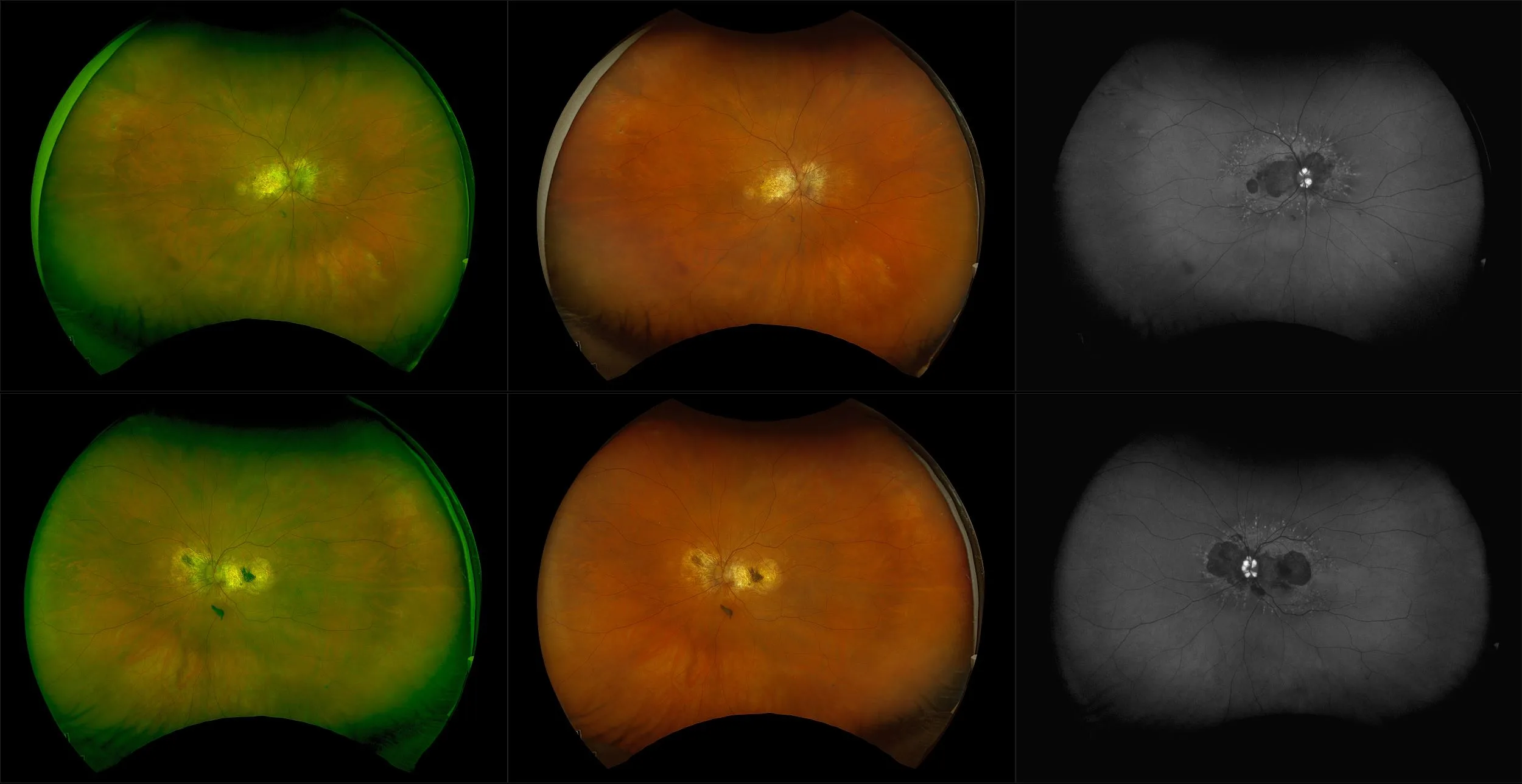
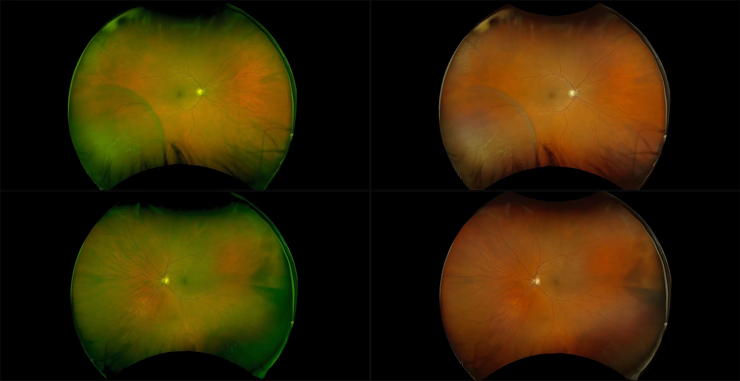
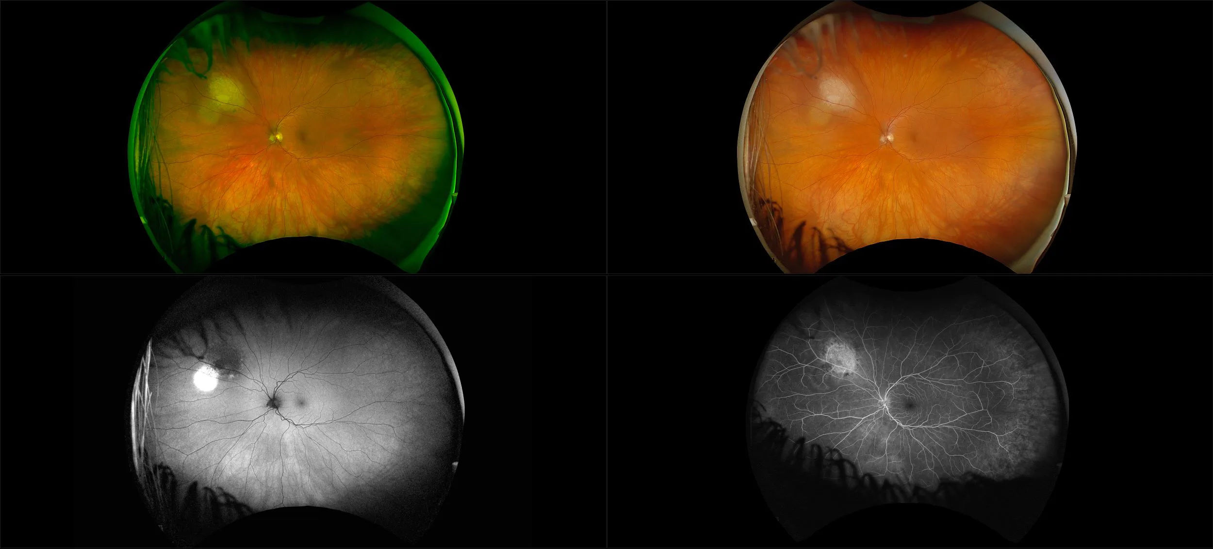
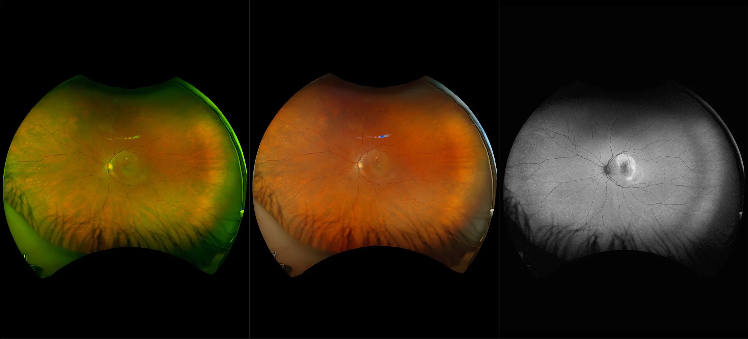
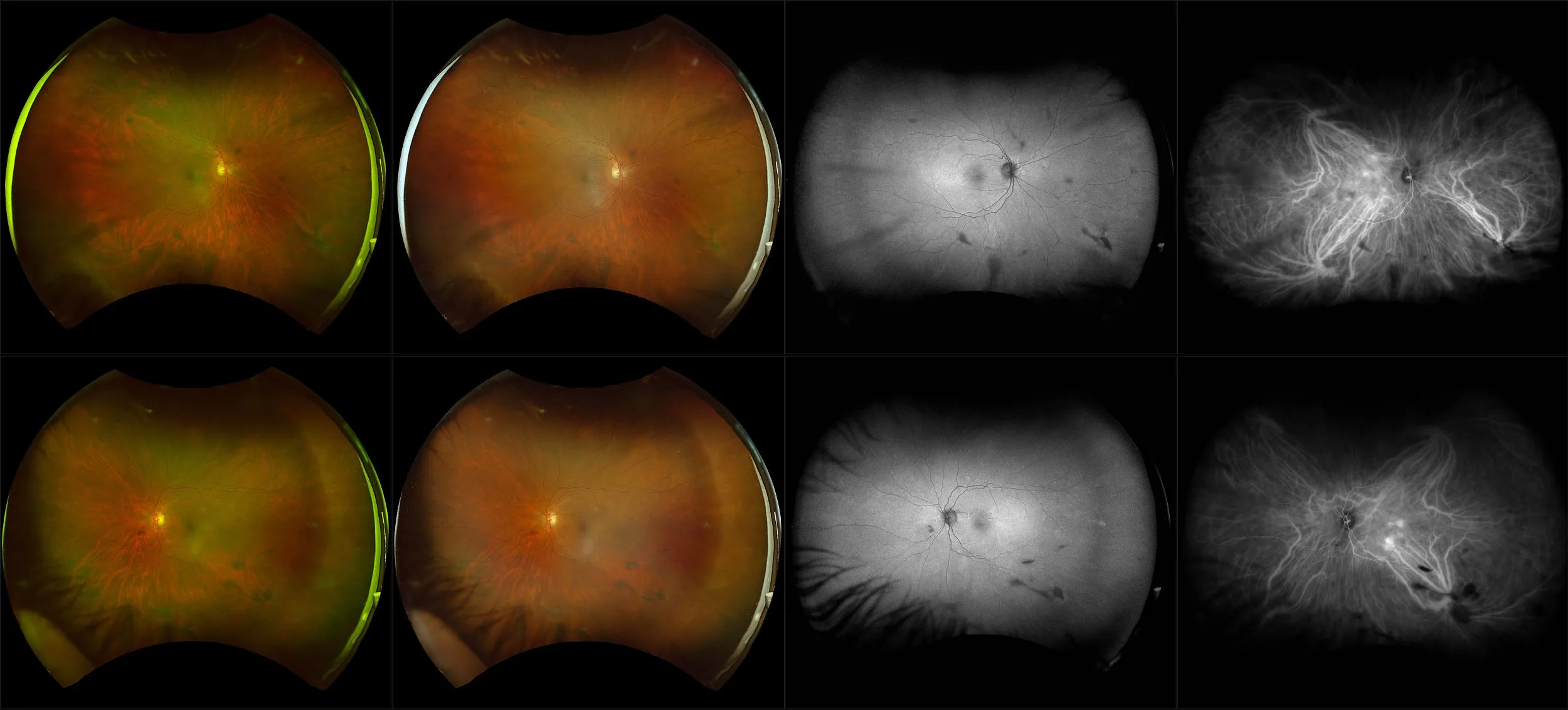
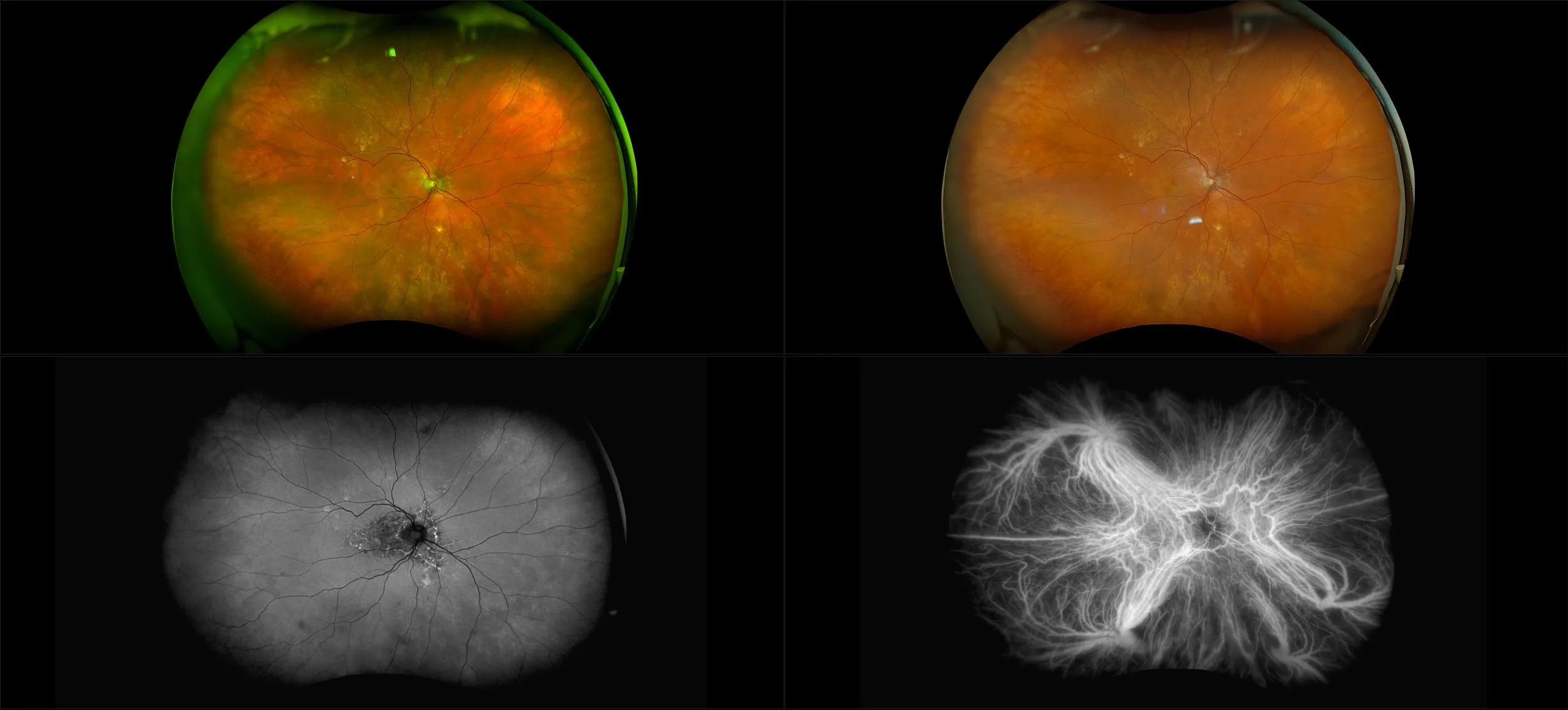
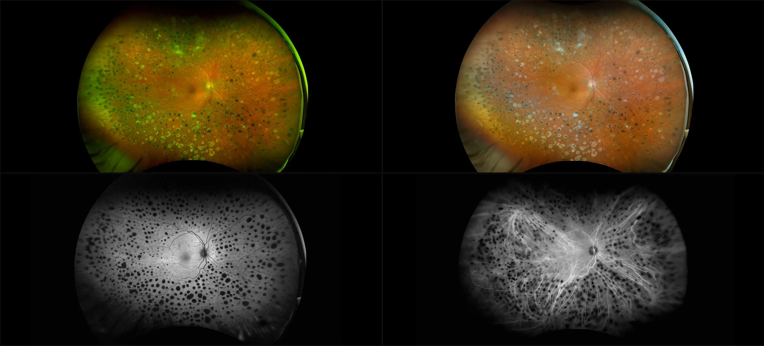
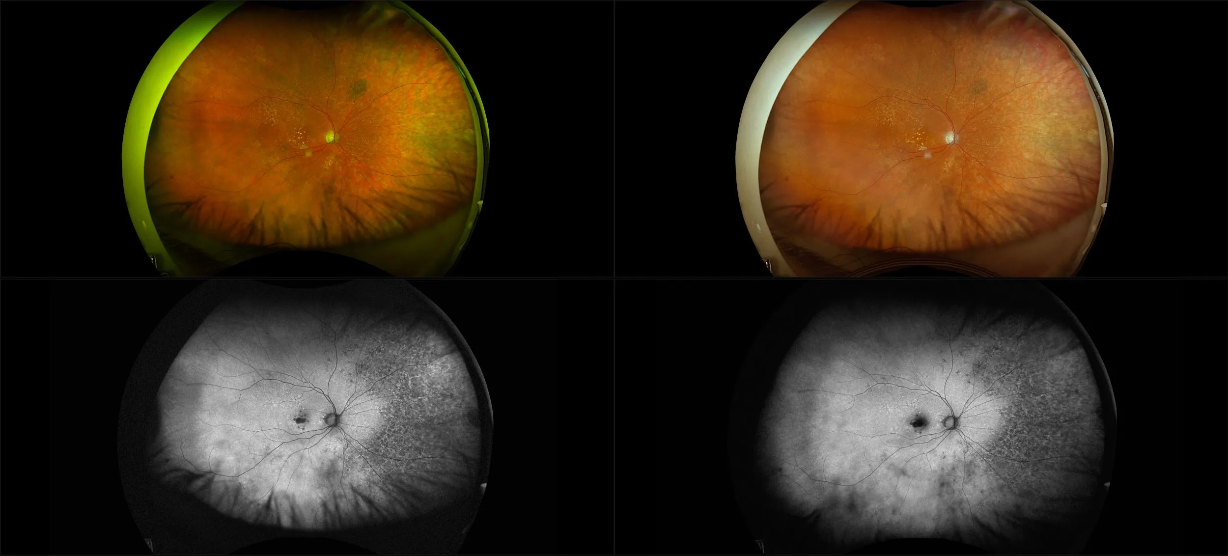
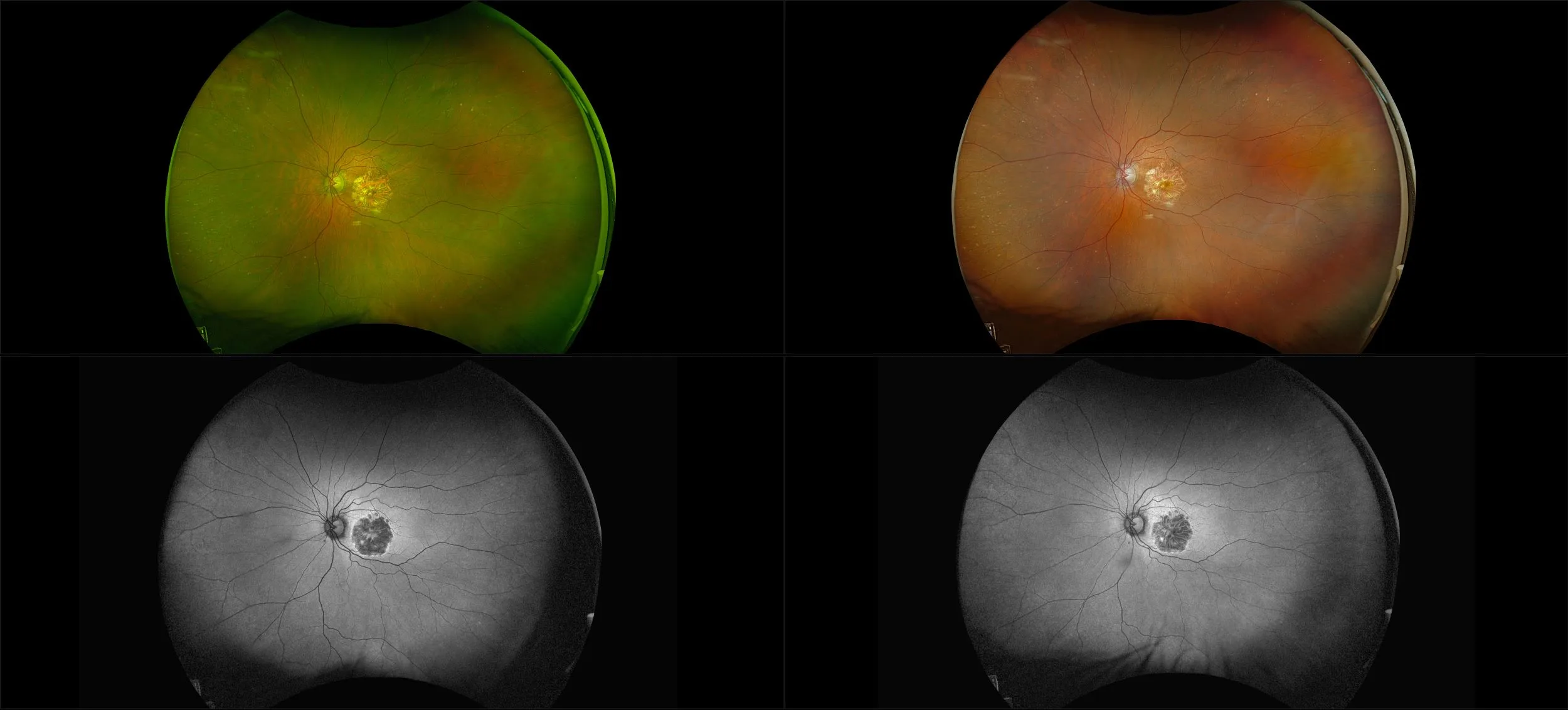
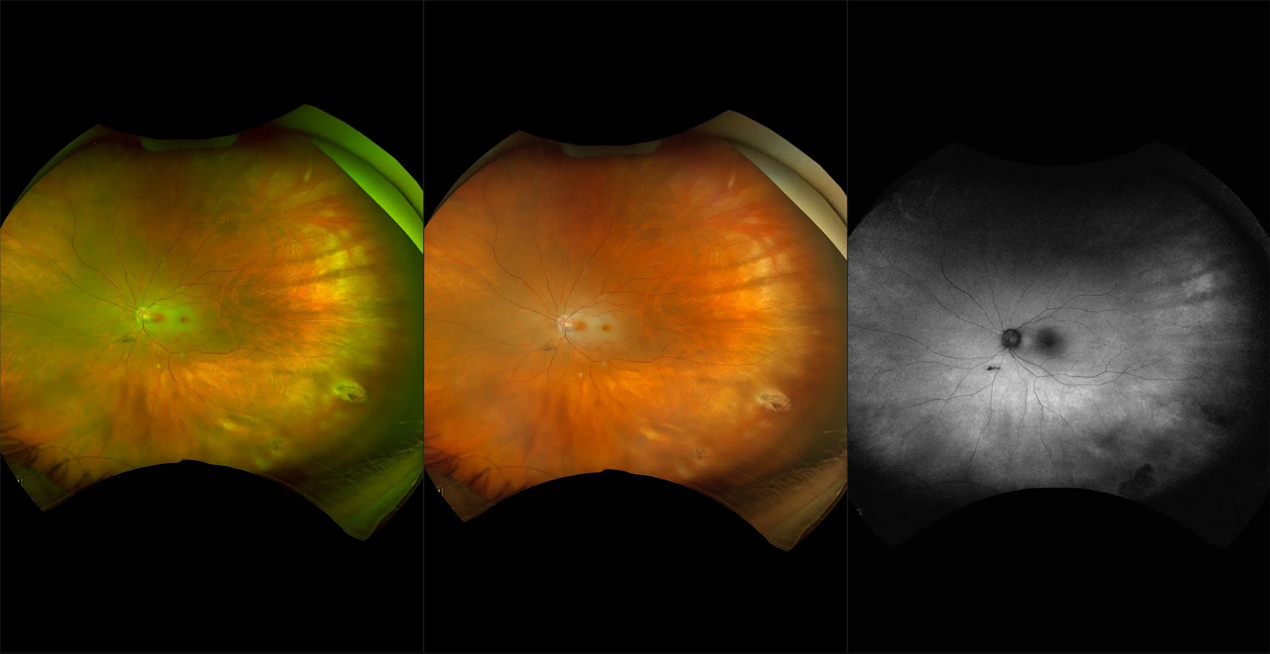
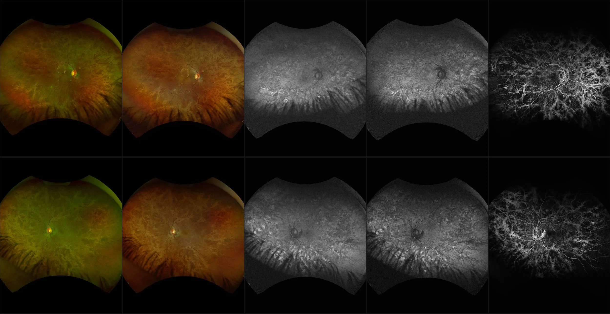
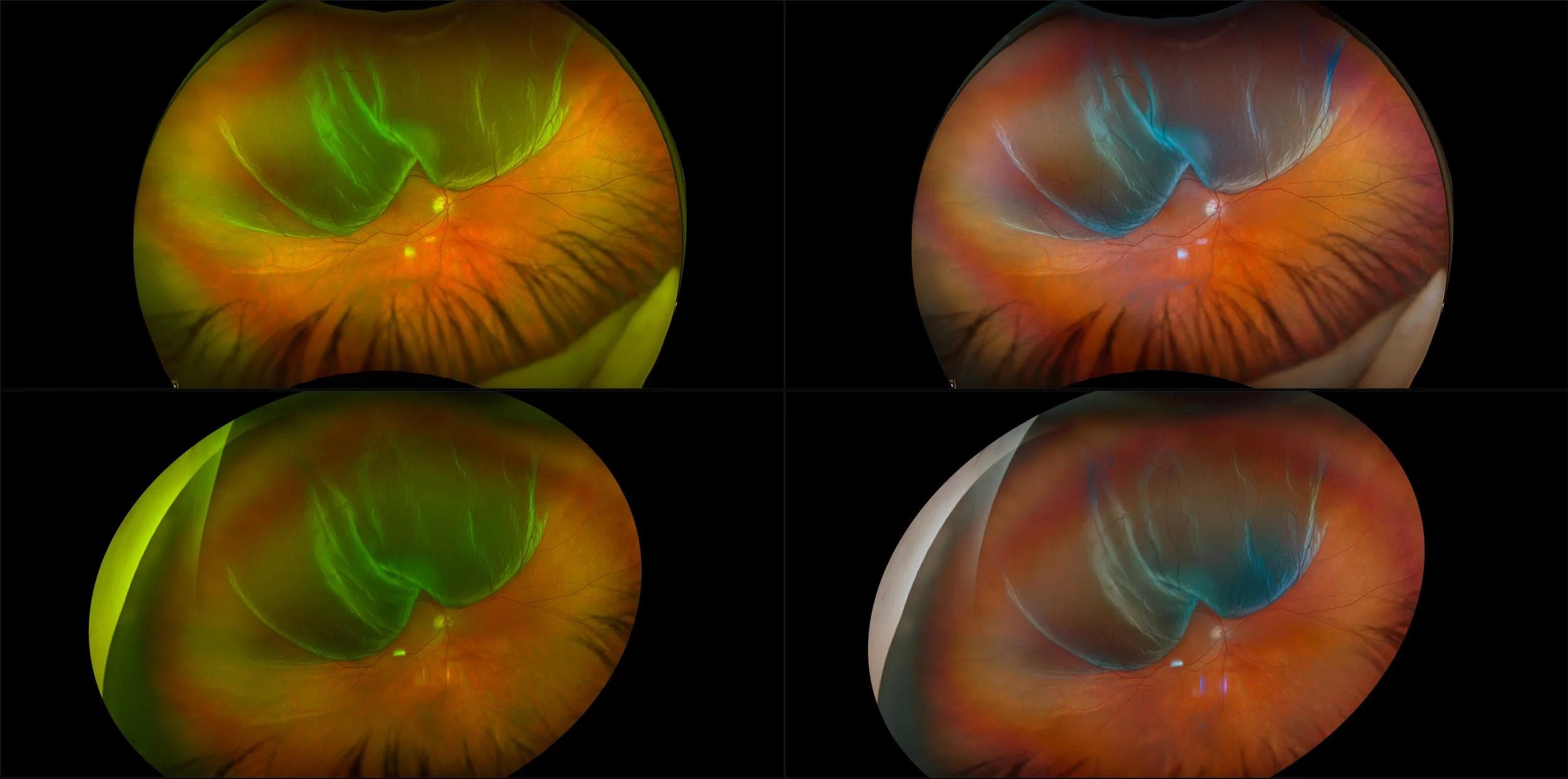
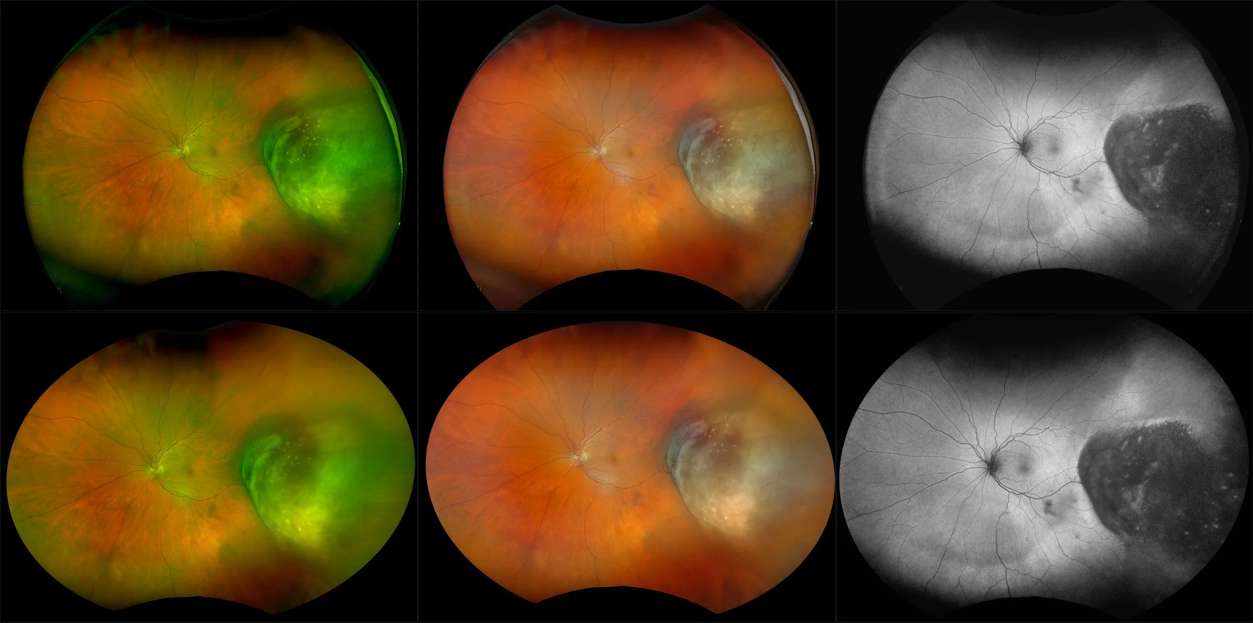
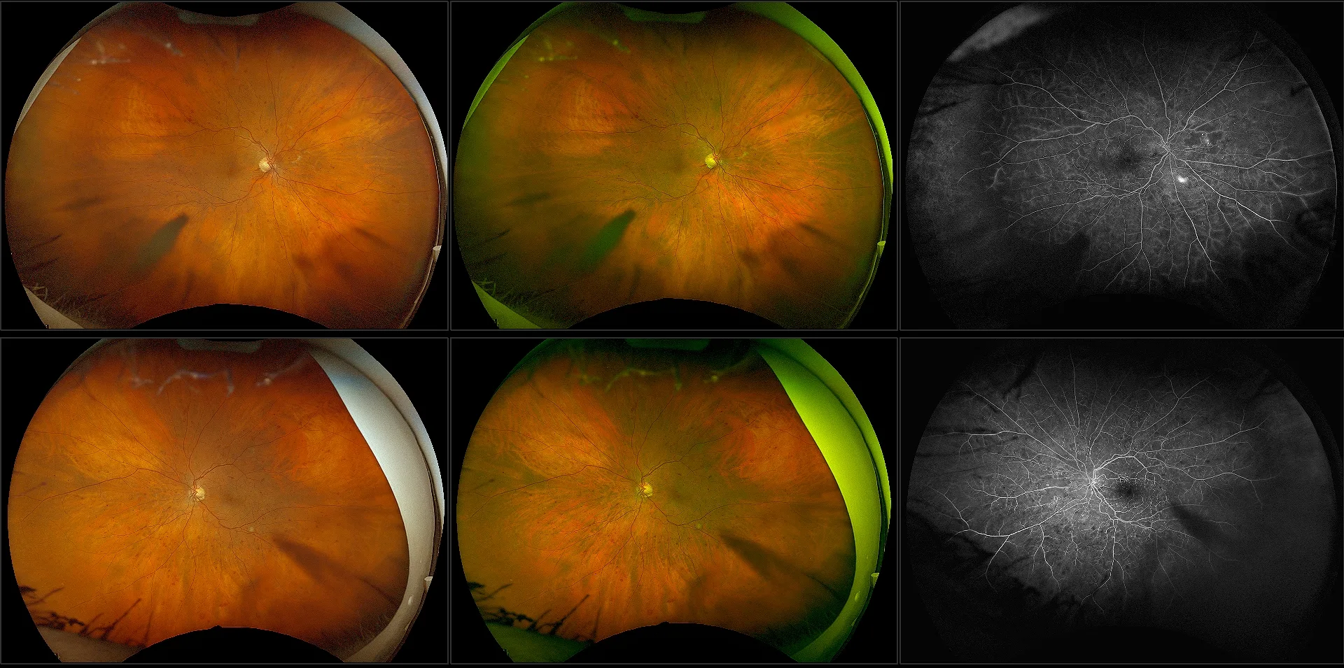
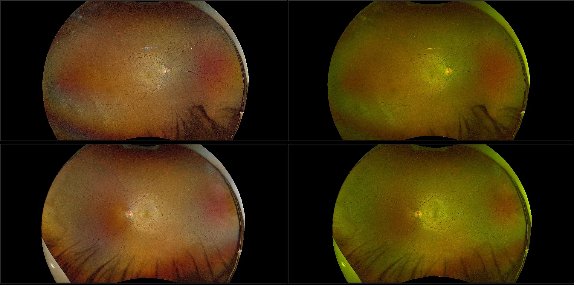
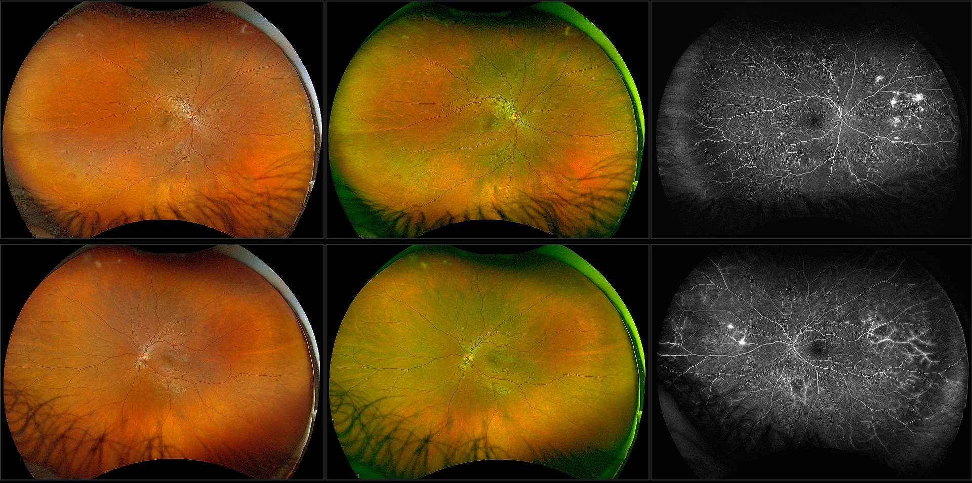
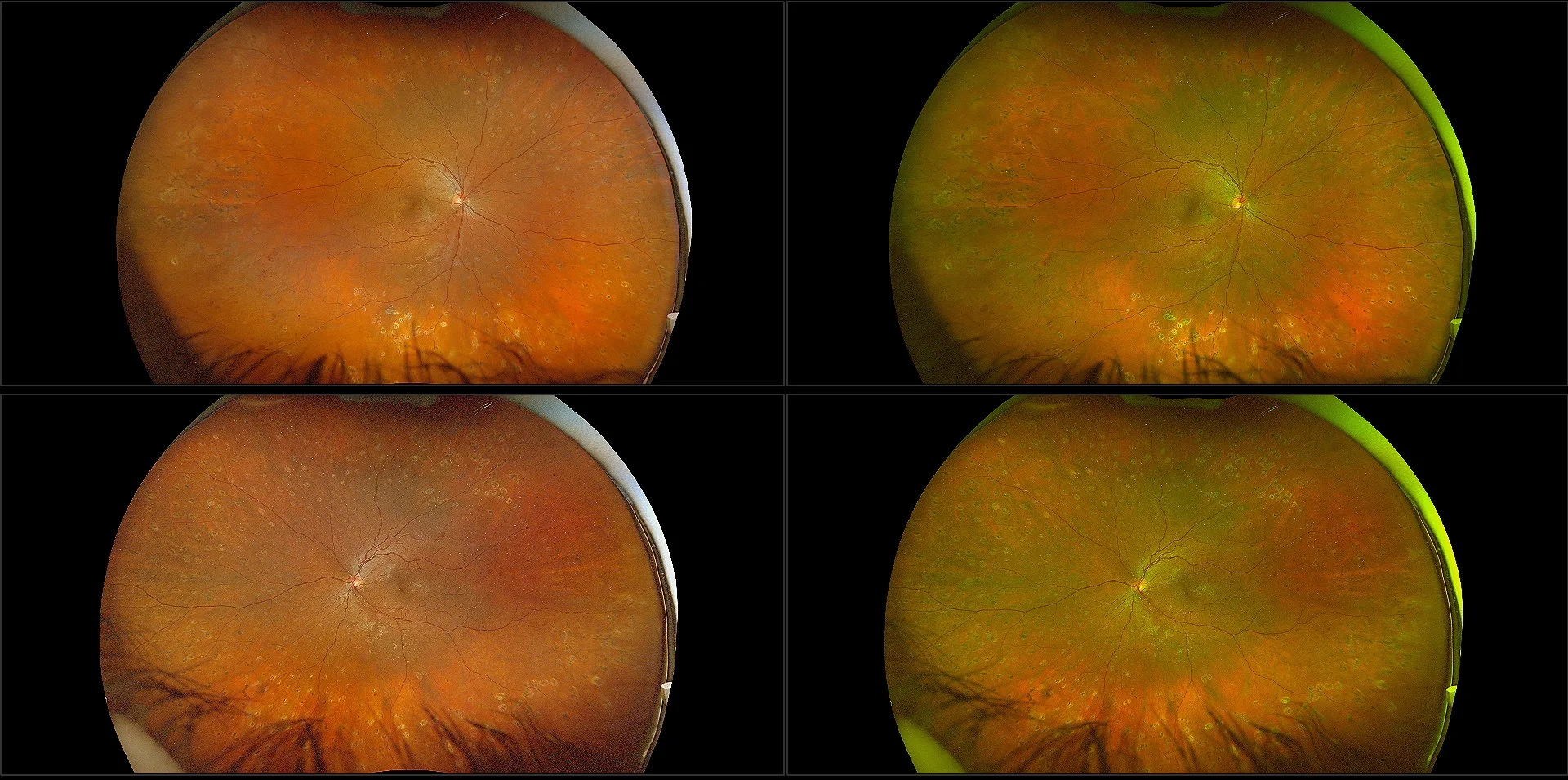
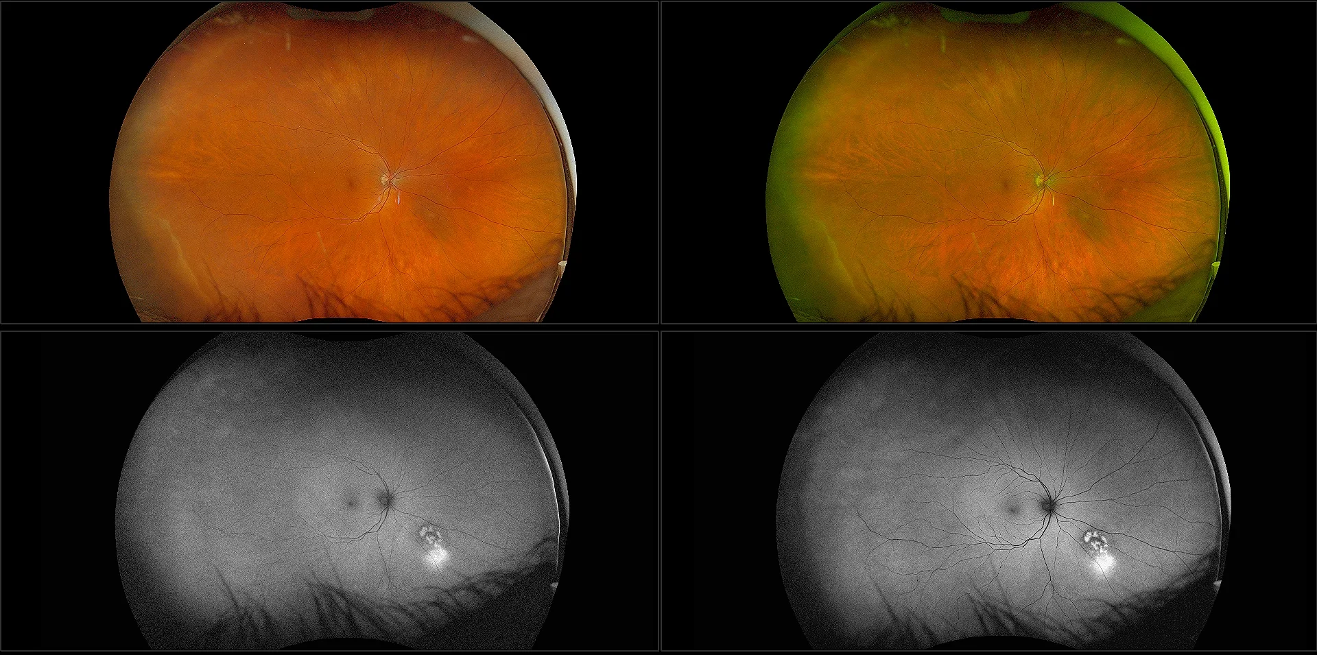
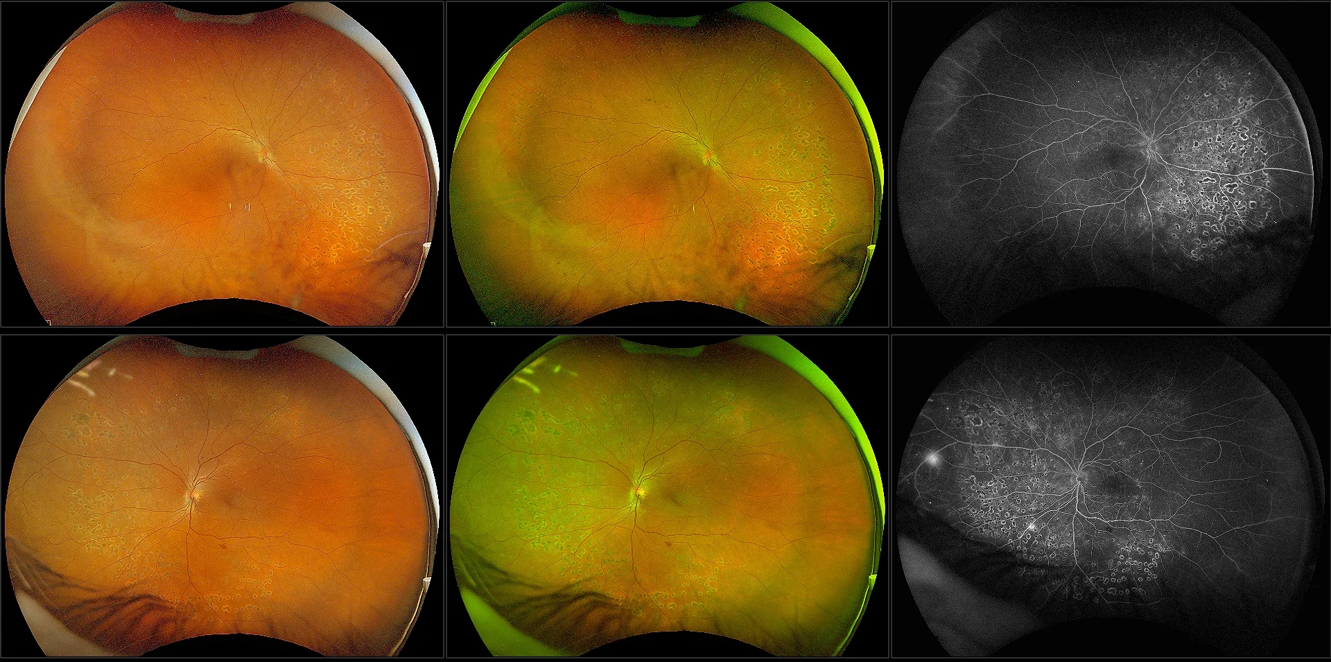
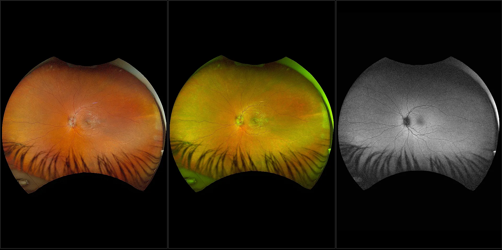
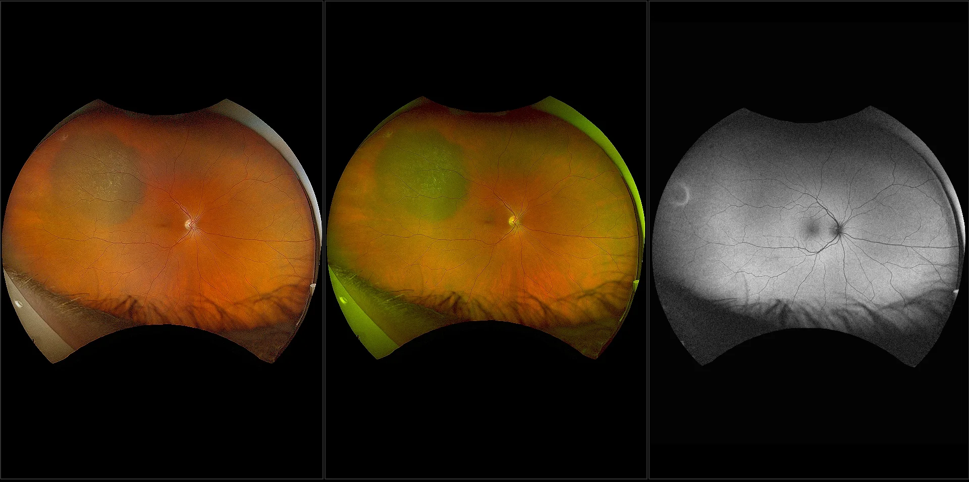
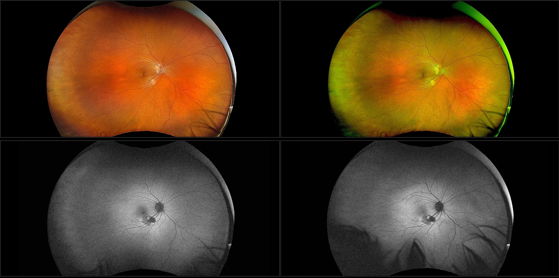
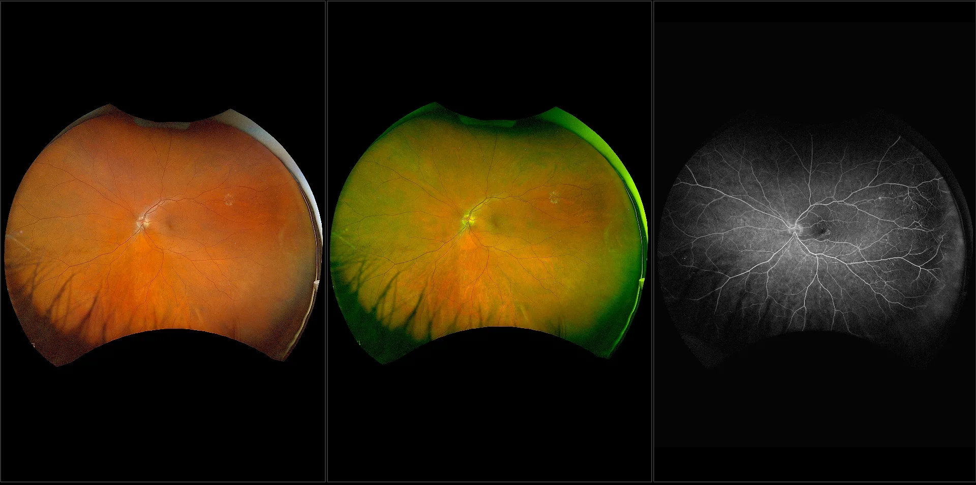
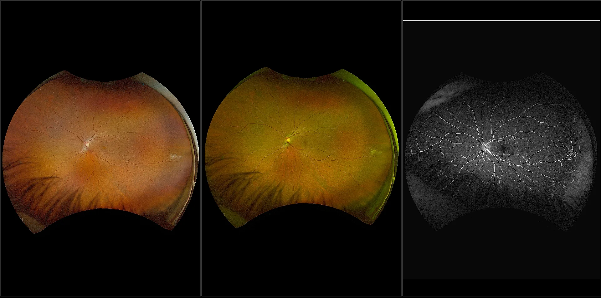
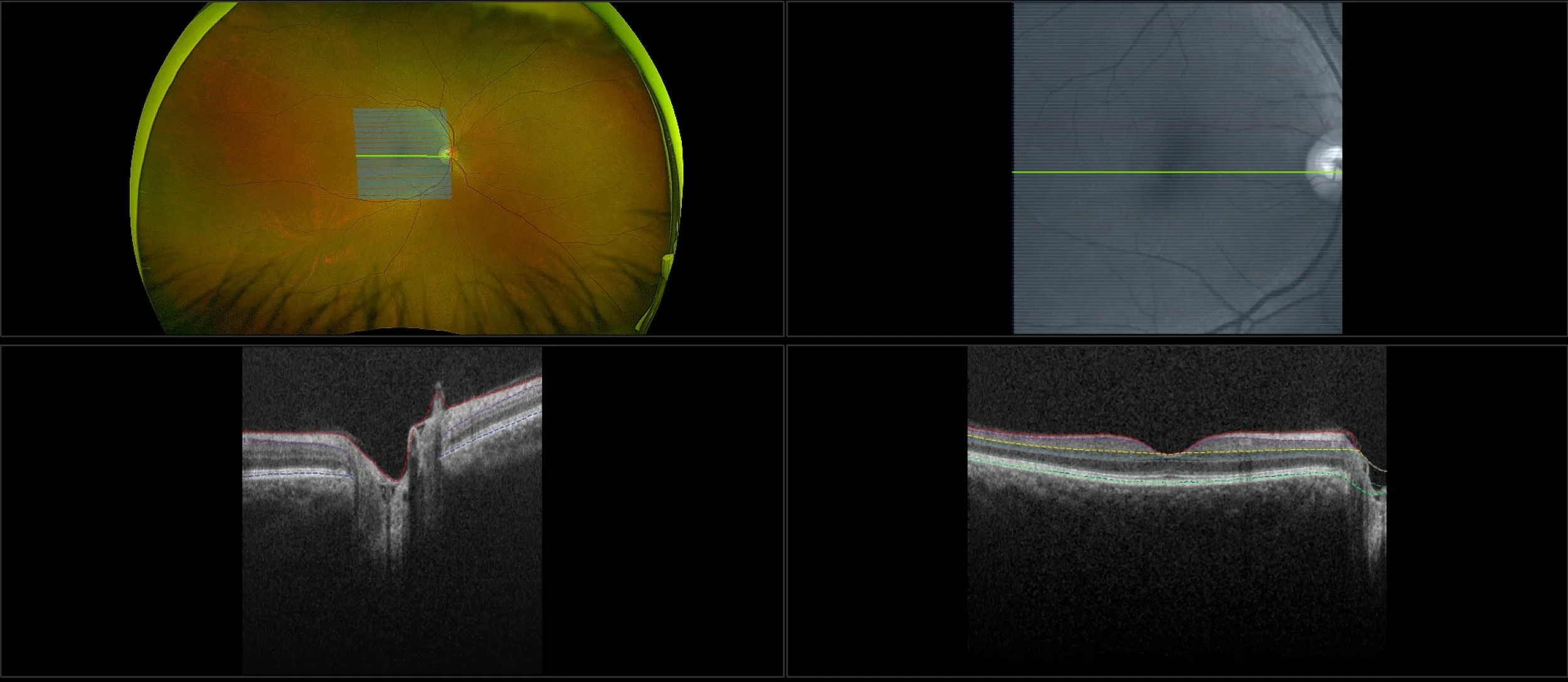
optomap Multimodal Color RGB Cases
Optos offers multimodal imaging with all ultra-widefield devices. Having both ultra-widefield and four images captured in less than one second has been shown to enhance pathology detection and disease management as well as improve practice and clinic flow. Ultra-widefield multimodal imaging is important across all access points of patient care - screening, detection, diagnosis, and treatment.
Related Cases


Additional Resources