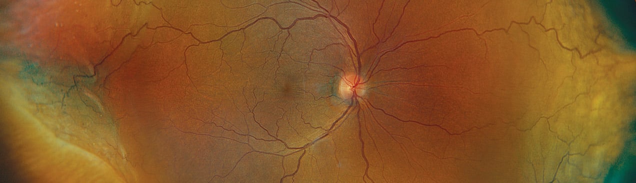
Defining Ultra-widefield (UWF™) Retinal Imaging
Optos technology has been installed in more than 30,000 locations globally and utilized in over 4,000 peer-reviewed papers, and 365+ disease states & pathologies. What differentiates widefield retinal imaging from ultra-widefield retinal imaging? In 2019, the International Widefield Study Group published their classification & guidelines on the definitions for widefield and UWF; they defined UWF as “images showing retinal anatomy anterior to the vortex vein ampullae in all four quadrants.” Optos technology represents the ONLY single capture retinal imaging that meets these criteria.
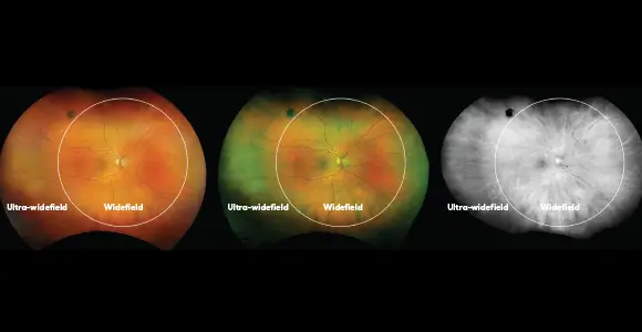
Widefield v Ultra-widefield v Optos Ultra-widefield v Pan-retinal
The International Widefield Imaging Study Group reviewed a set of images from various manufacturers to help support the generation of the following definitions when describing the field of view of retinal images1:
Widefield – centered on the fovea and includes the retina in all four quadrants posterior to and including the vortex vein ampullae.
Ultra-widefield – images showing retinal anatomy anterior to the vortex vein ampullae in all four quadrants – Optos UWF does this in a single, 200⁰ image capture in less than ½ second, whereas the other products require multiple images and increased time to be considered ultra-widefield.
Pan-retinal – ora-to-ora image of the retina either in the horizontal or vertical direction.
optomap Increases Patient Workflow
In this study and other studies combined, optomap imaging has been demonstrated to capture the widest field of view in a single capture of any imaging technology: more than 50% additional retinal area captured versus one single-capture widefield lens based system2, 110° more than a single capture widefield image and 65° more than the montaged ultra-widefield image from another lens based camera system3, more vortex veins visualized4 and statistically significantly more retinal surface area.5,6
“A single capture image which provides a view of the vortex veins in all four quadrants and beyond, thus meeting the widefield & ultra-widefield definitions, would offer enhanced efficiency in a real-world clinical setting versus a montage image, whether it be manual or automated.”
We encourage you to contact us if you have questions regarding how optomap and Optos technology can benefit your patients and clinical settings.
Clinical Summary
optomap Defining Ultra-widefield
optomap is the only single shot UWF retinal image, by definition.
Referenced Papers
Classification and Guidelines for Widefield Imaging Recommendations from the International Widefield Imaging Study Group.
Potential efficiency benefits of nonmydriatic ultrawide field retinal imaging in an ocular telehealth diabetic retinopathy program.
Identification of Diabetic Retinopathy and Ungradable Image Rate with Ultrawide Field Imaging in a National Teleophthalmology Program - Ophthalmology
A novel hybrid fixed and mobile ultrawide field imaging program for diabetic tele-retinopathy screening.
Comparison of Early Treatment Diabetic Retinopathy Study Standard 7-Field Imaging With Ultrawide-Field Imaging for Determining Severity of Diabetic Retinopathy
Peripheral Lesions Identified on Ultrawide Field Imaging Predict Increased Risk of Diabetic Retinopathy Progression over 4 Years.
Peripheral Retinal Changes Associated with Age-Related Macular Degeneration in the Age-Related Eye Disease Study 2.
Area of peripheral retinal nonperfusion and treatment response in branch and central retinal vein occlusion.
The area of peripheral retinal nonperfusion is variable in patients with retinal vein occlusion and affects its clinical course and response to treatment.