Feasibility and Clinical Utility of
Ultra-Widefield Indocyanine
Green Angiography
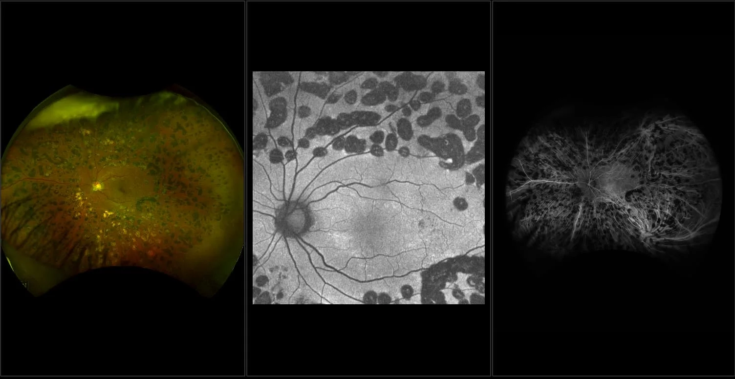
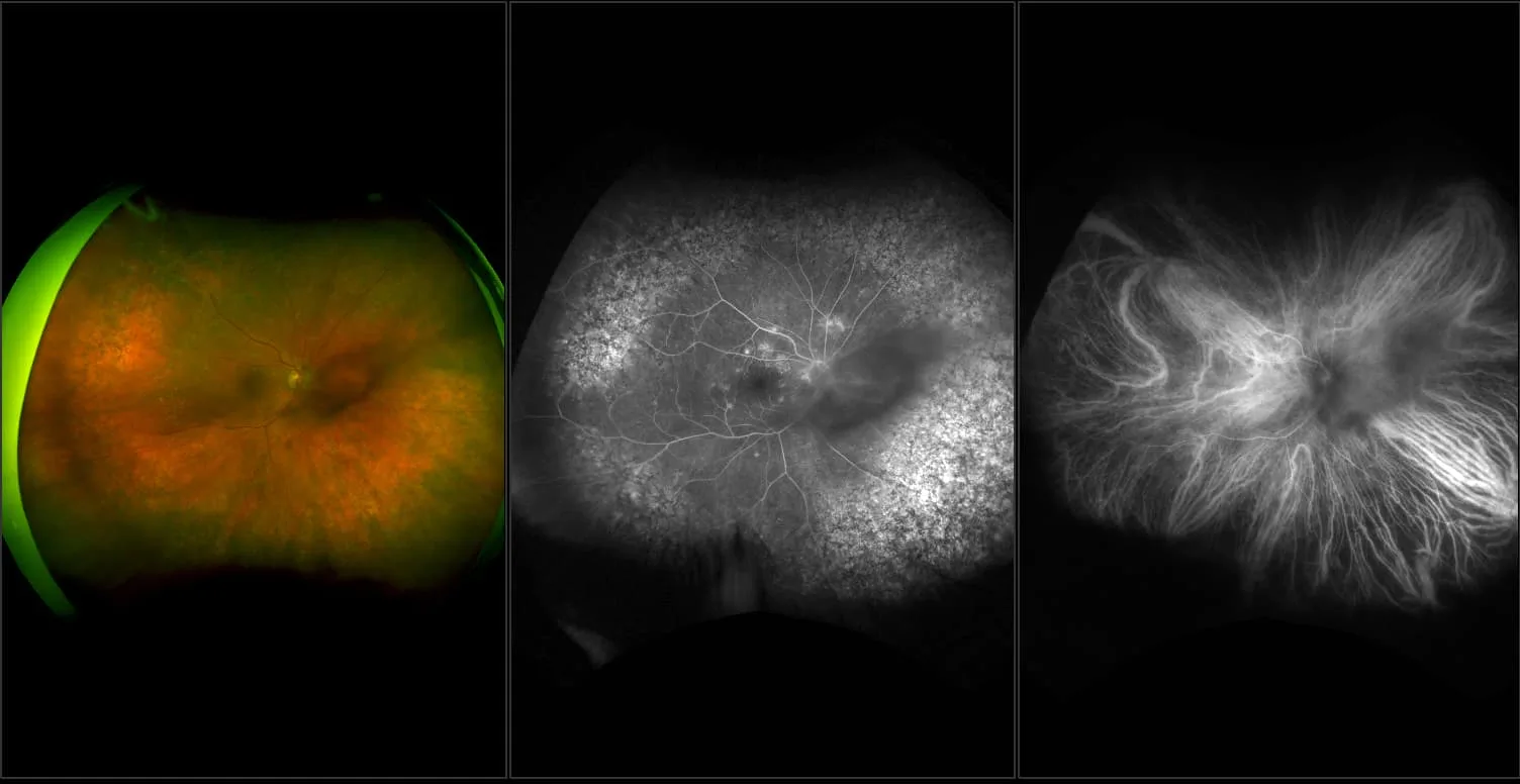
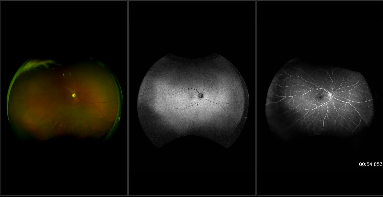
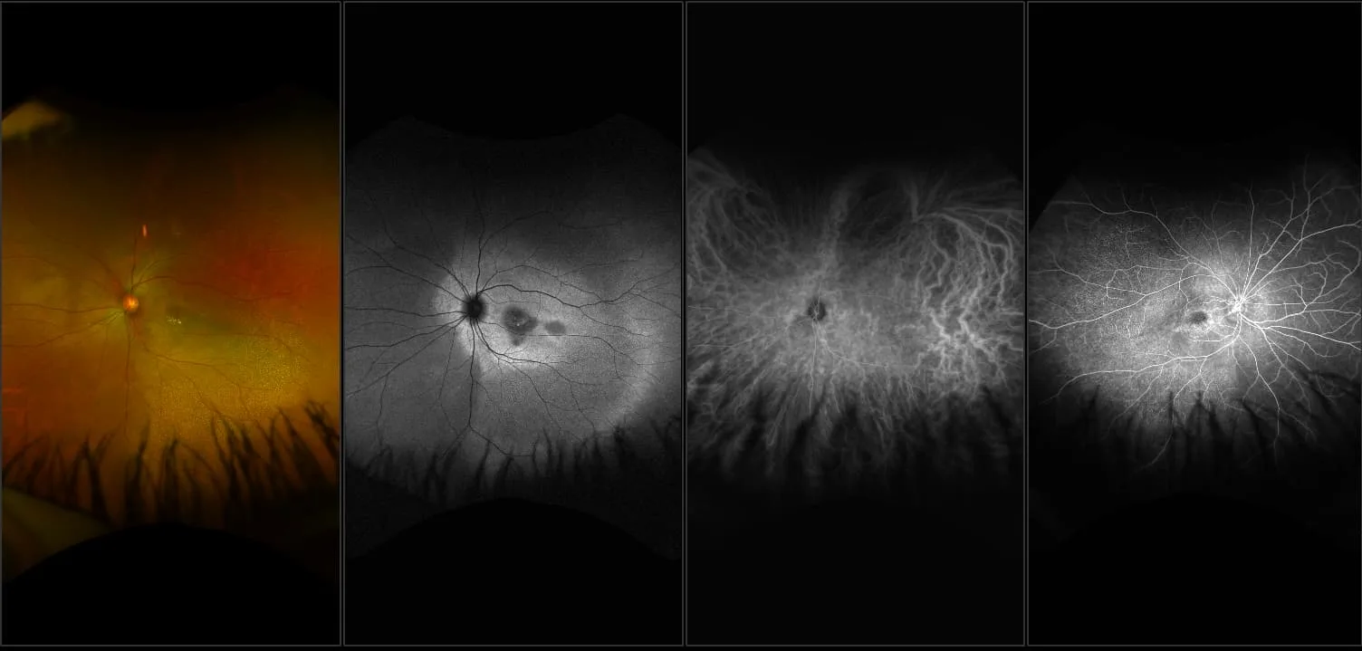
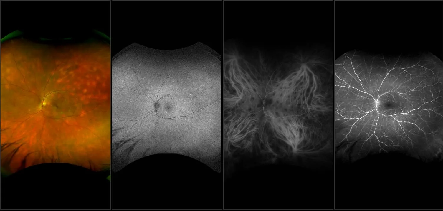
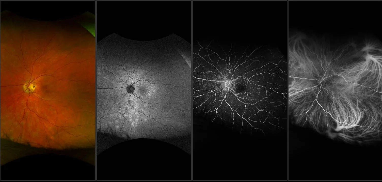
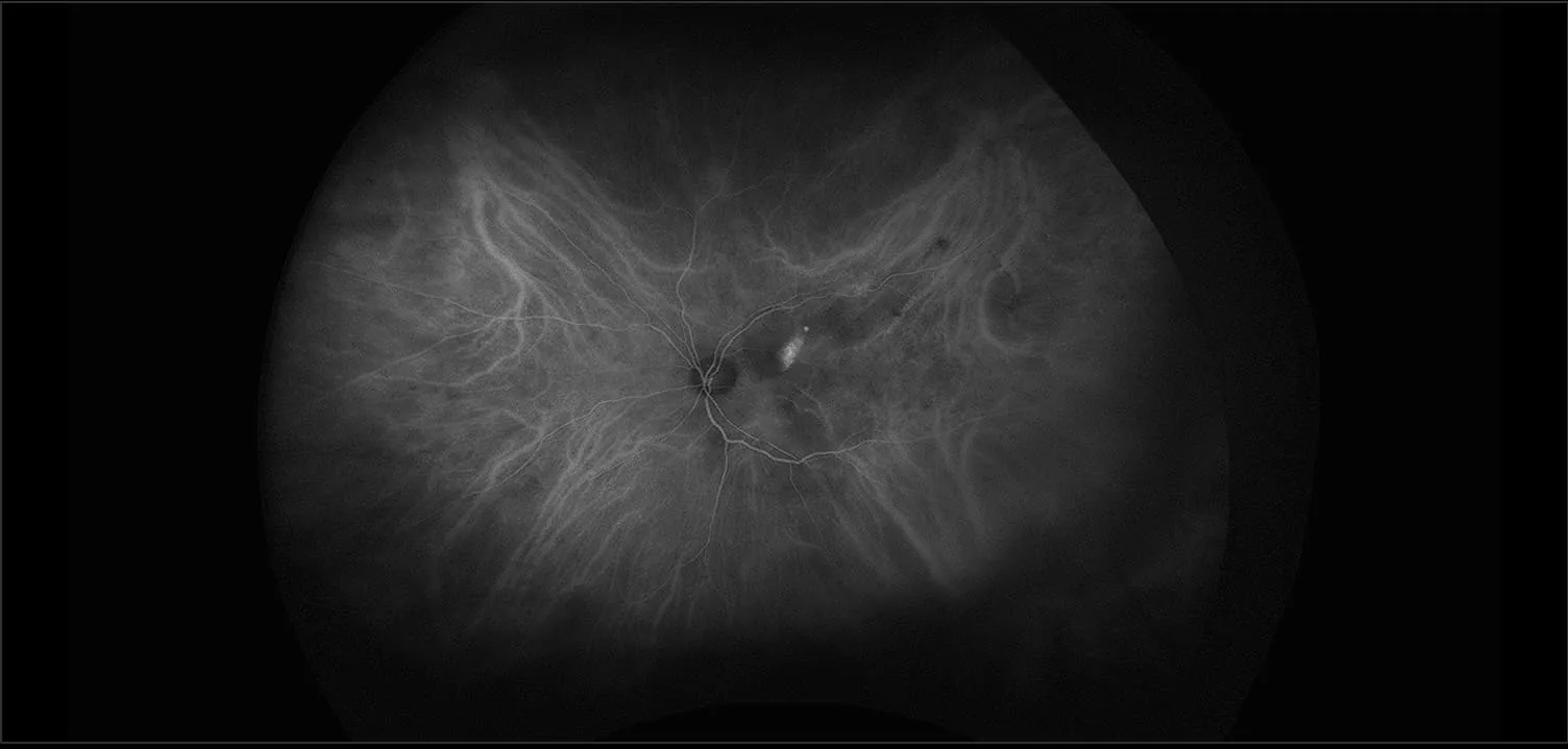
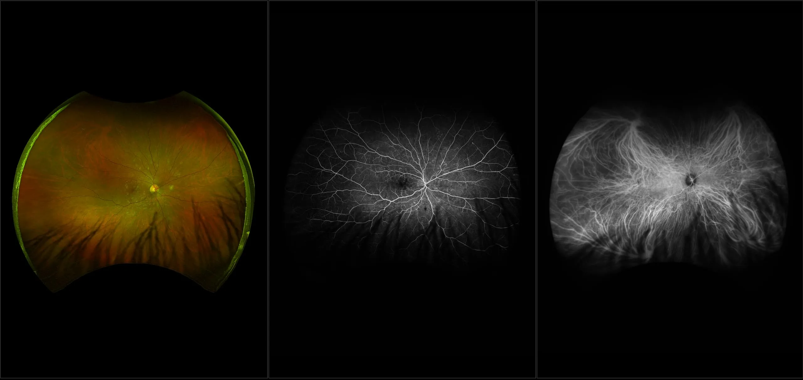
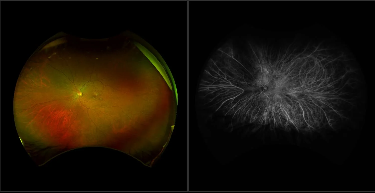
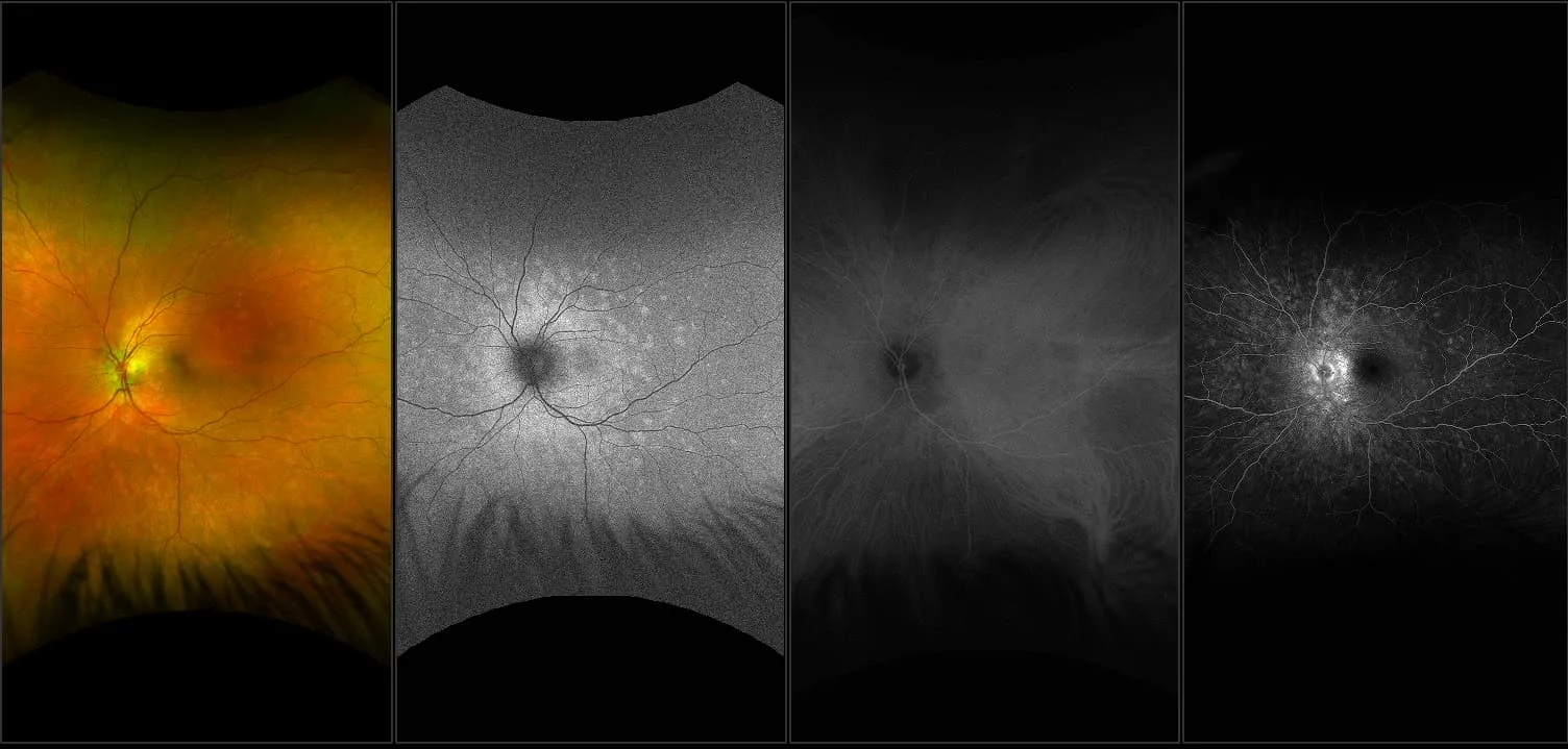
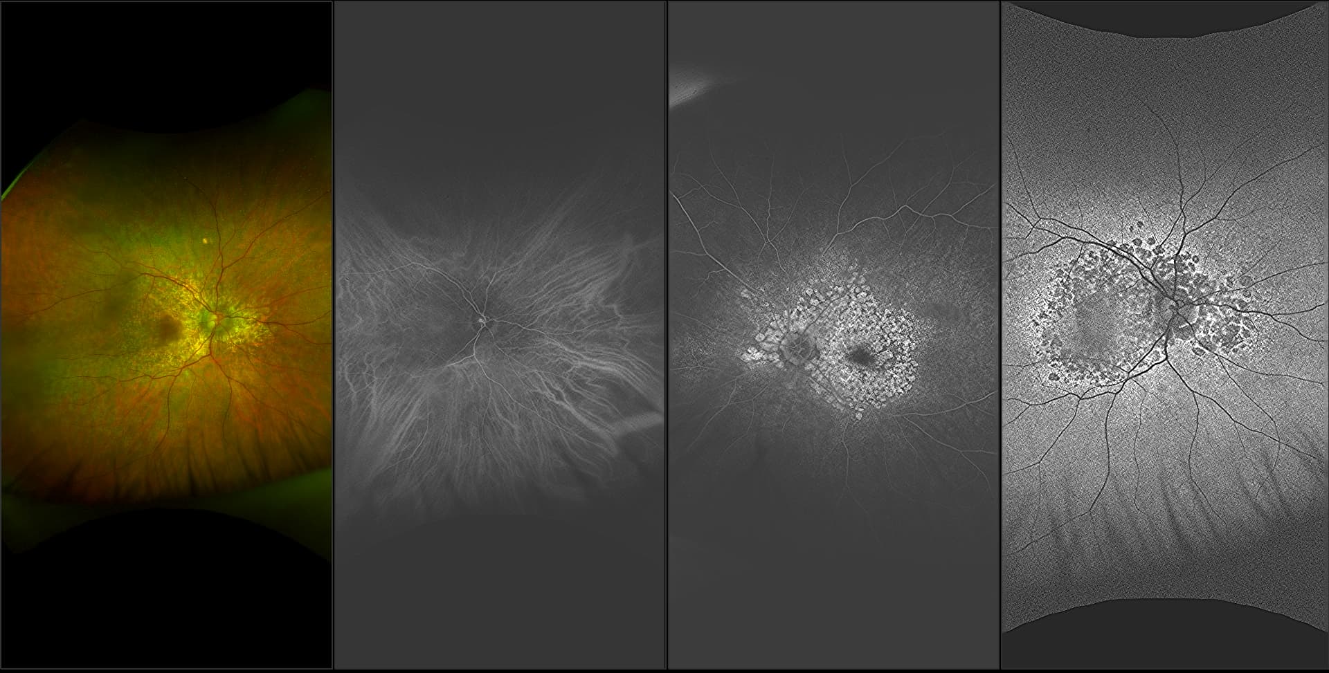
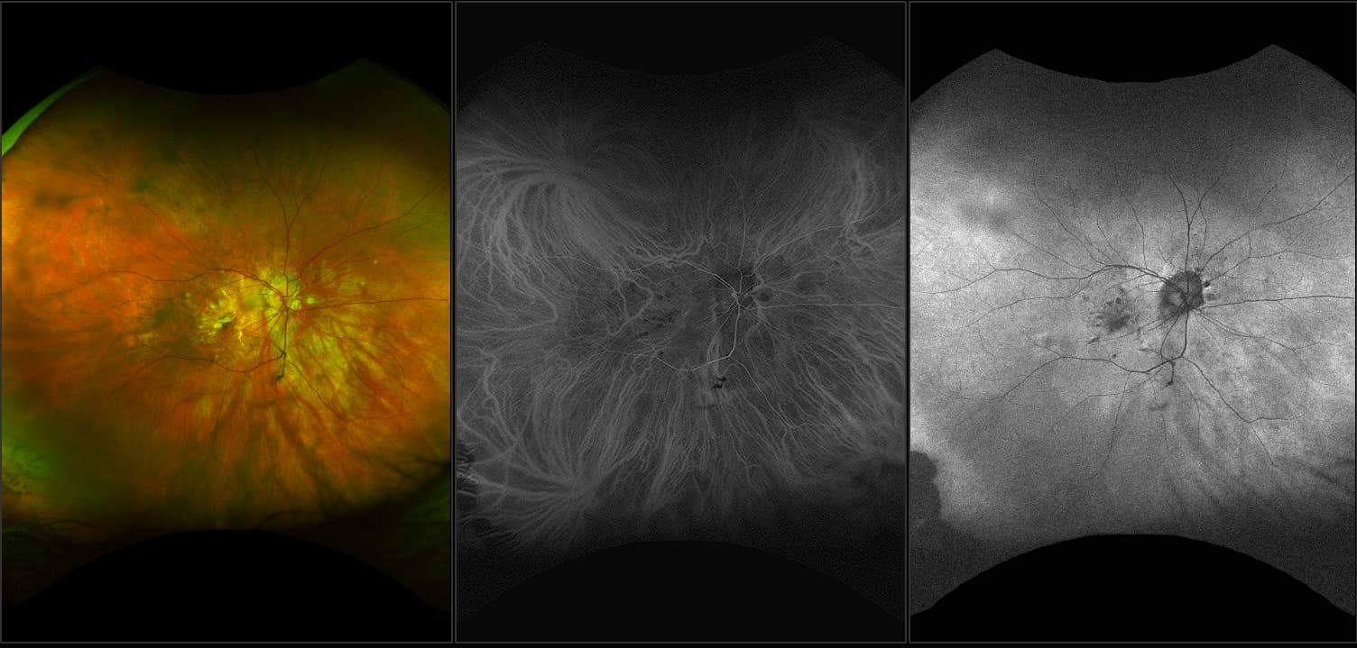
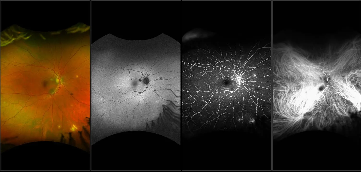
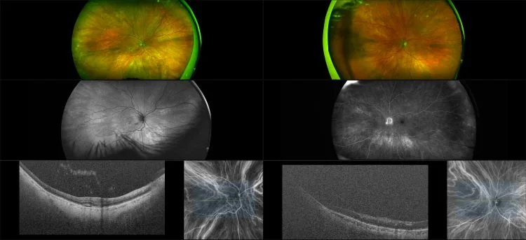
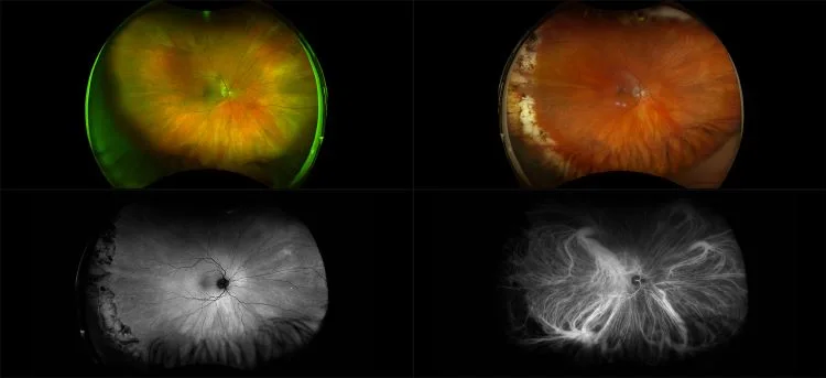
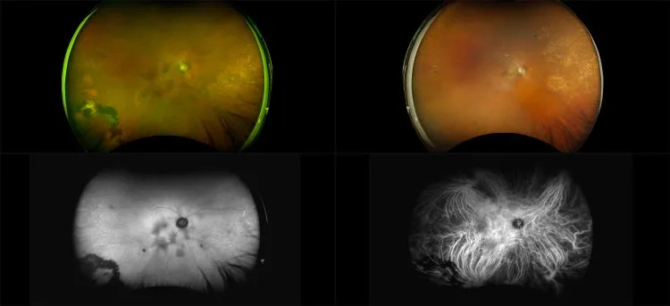
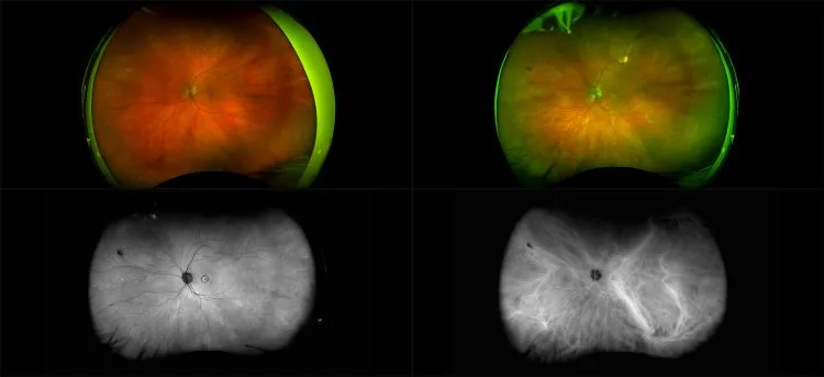
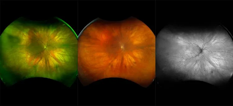
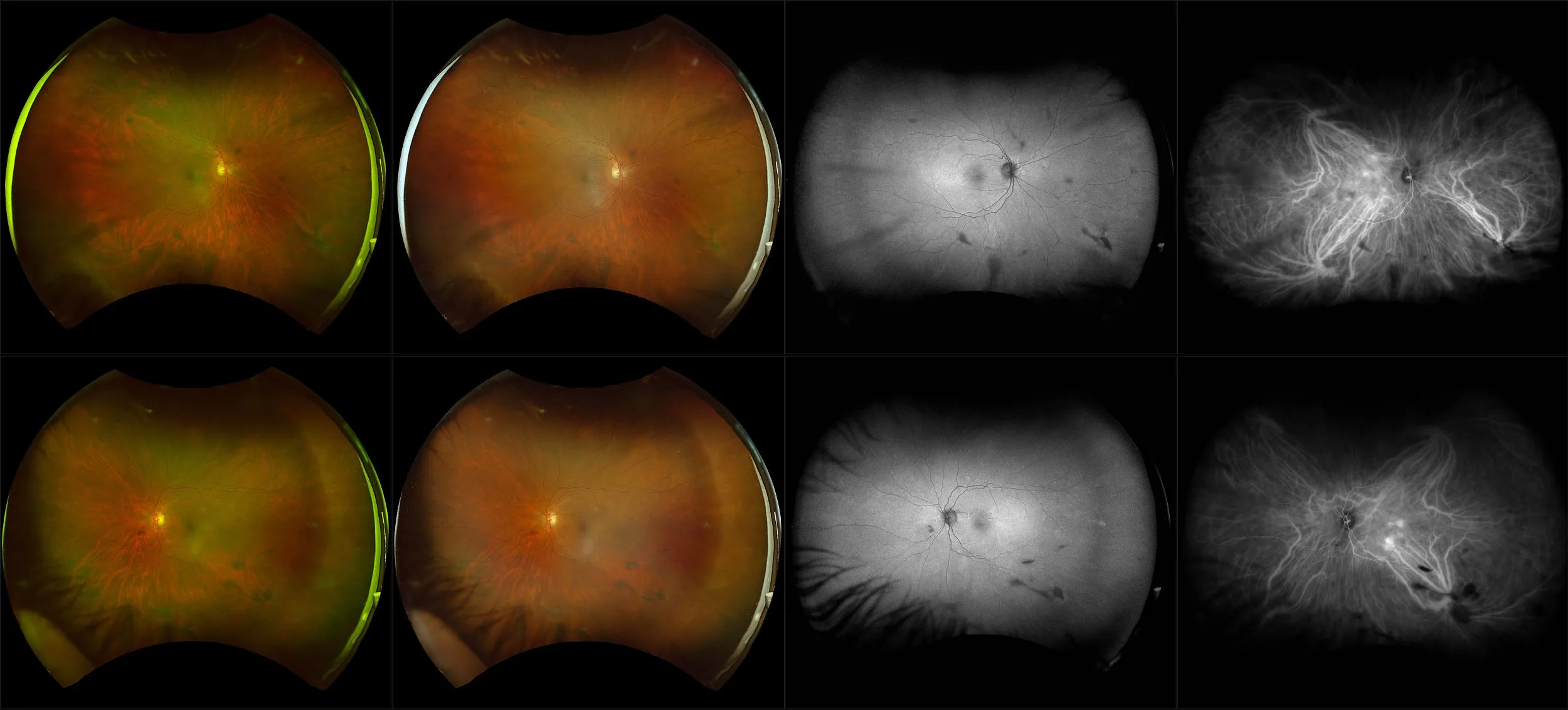
optomap icg ultra-widefield retinal imaging provides a more comprehensive view of the retina and choroid, revealing findings that conventional small-field imaging may miss. Clinical evidence shows that 67% of eyes present with peripheral changes detectable through this advanced approach, offering eye care professionals valuable insights for earlier detection and improved patient management. With its ability to extend visibility into the periphery, optomap ICG enhances diagnostic confidence and supports better decision-making in clinical practice.
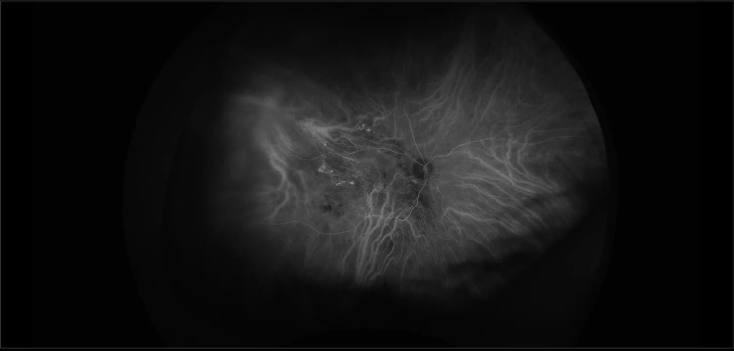
“Ultra-widefield indocyanine angiography reveals abnormalities in the peripheral retina that may otherwise be missed on conventional ICGA imaging.”1
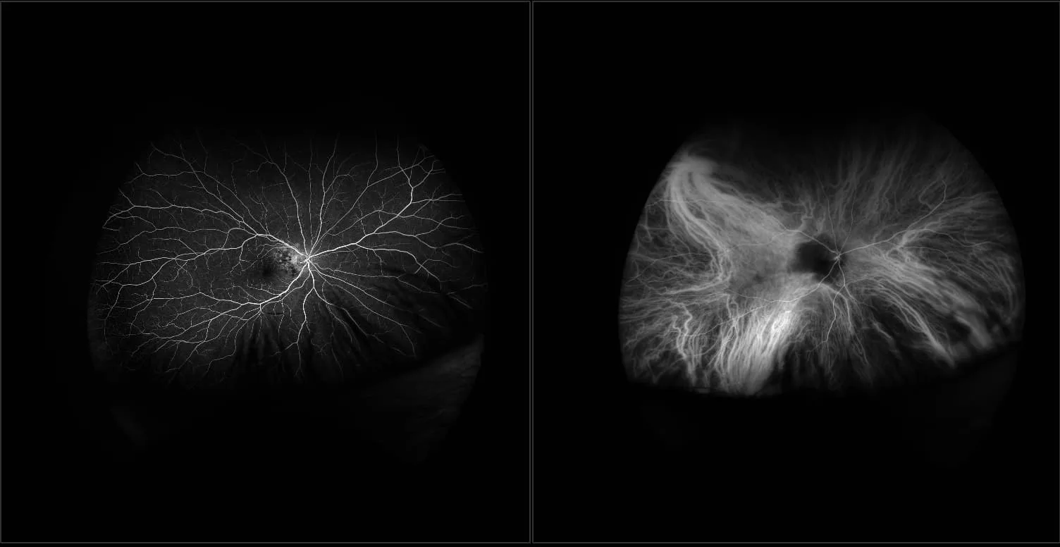
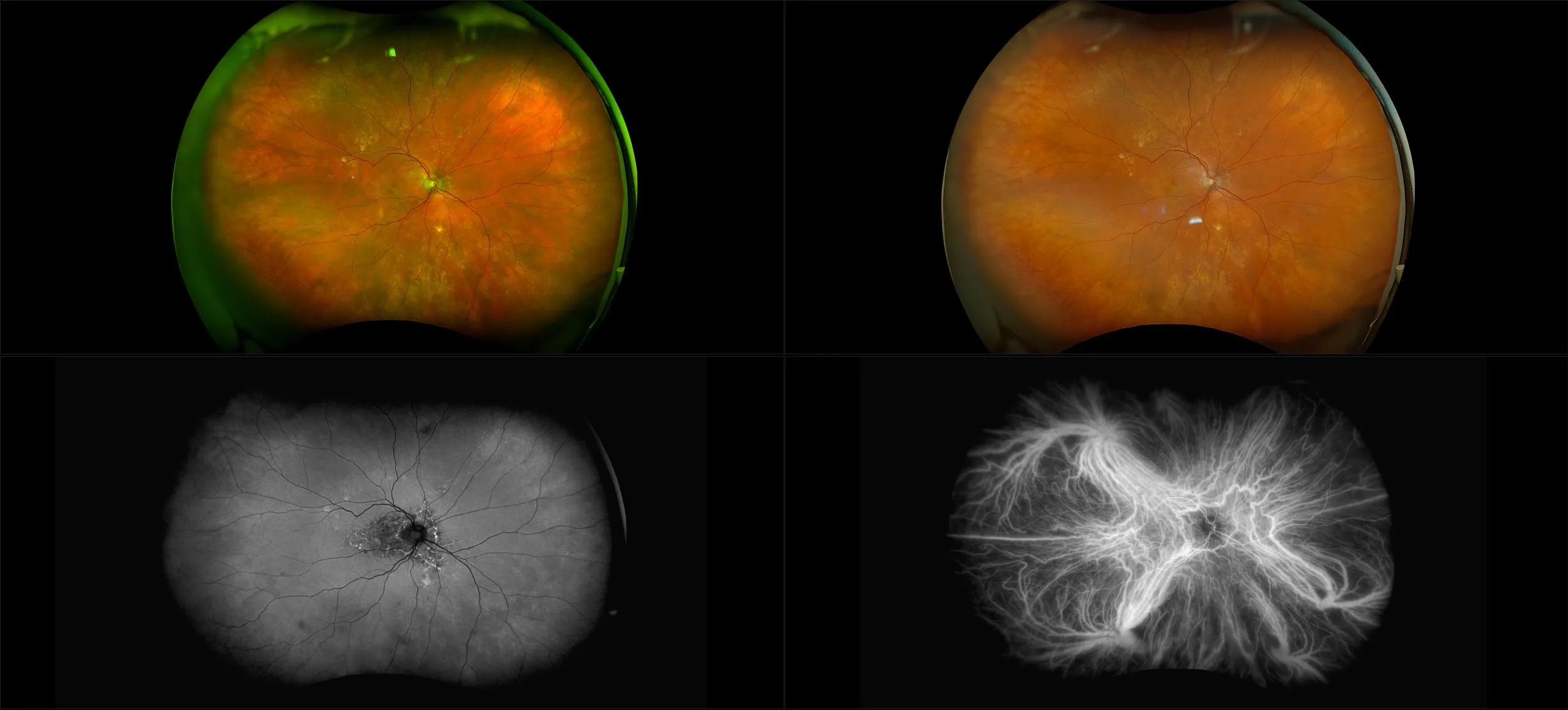
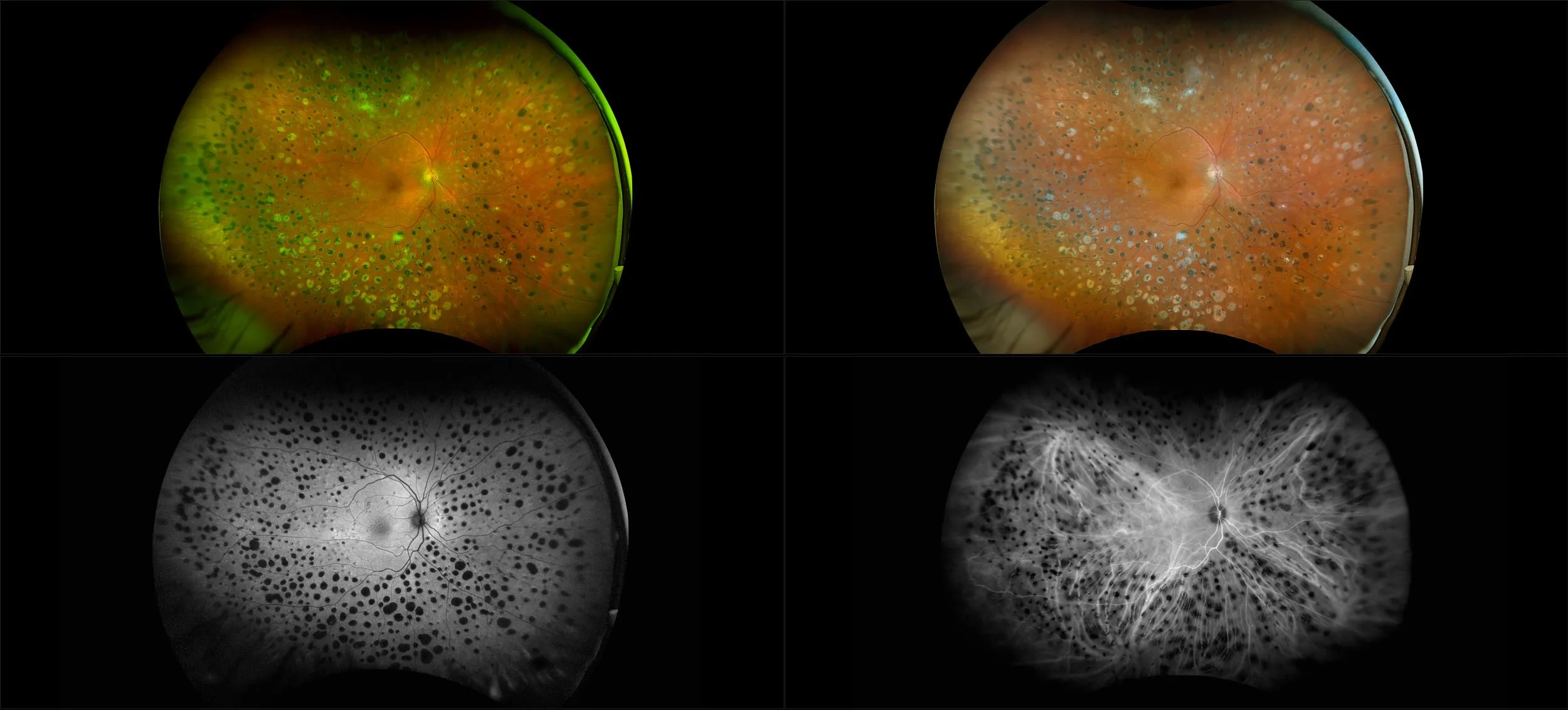
optomap icg is redefining the understanding of the choroid, in a study of normals, the mean number of vortex vein ampullae is much larger than previously reported, with a mean number of 8 with as many as 13.2 The normal peripheral extent of choroidal circulation on optomap icg was estimated to be 893.22mm.2,3 optomap icg also captured significant peripheral changes in 80% of AMD patients1 further underlining the importance of using ultra-widefield imaging for ICG evaluation.
Clinical Summary
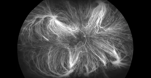
67% of Eyes have Peripheral Findings on optomap ICG
UWF icg reveals abnormalities in the peripheral retina that may be missed on conventional icga imaging.
Referenced Papers
Feasibility and Clinical Utility of Ultra-Widefield Indocyanine Green Angiography.
Distribution and Location of Vortex Vein Ampullae in Healthy Human Eyes as Assessed by Ultra-Widefield Indocyanine Green Angiography.
Clinical utility of ultra-widefield indocyanine green angiography in posterior uveitis
Ultra-wide field indocyanine green angiography in central serous chorioretinopathy
Peripheral extent of the choroidal circulation by ultra-widefield indocyanine green angiography in healthy eyes.
Classification and Guidelines for Widefield Imaging Recommendations from the International Widefield Imaging Study Group.
Choroidal Vascular Abnormalities by Ultra-widefield Indocyanine Green Angiography in Polypoidal Choroidal Vasculopathy.