optomap is More Informative and
Cost-effective for Ocular Oncology
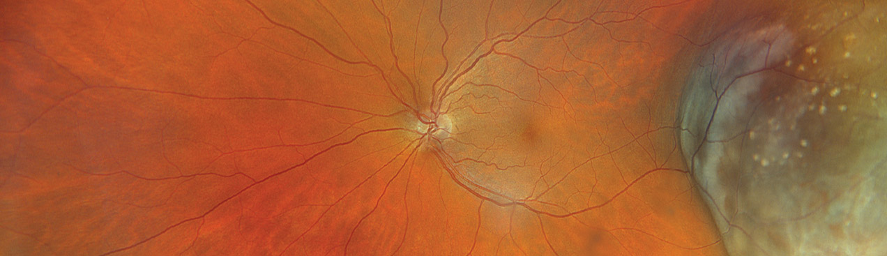
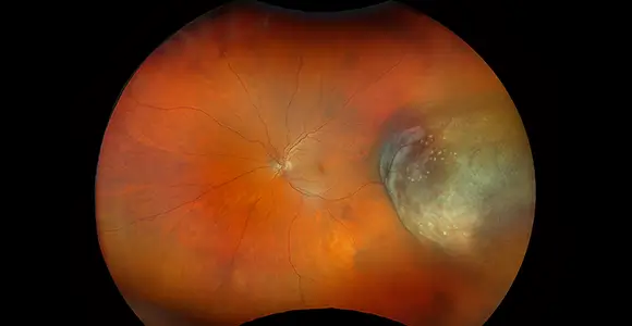
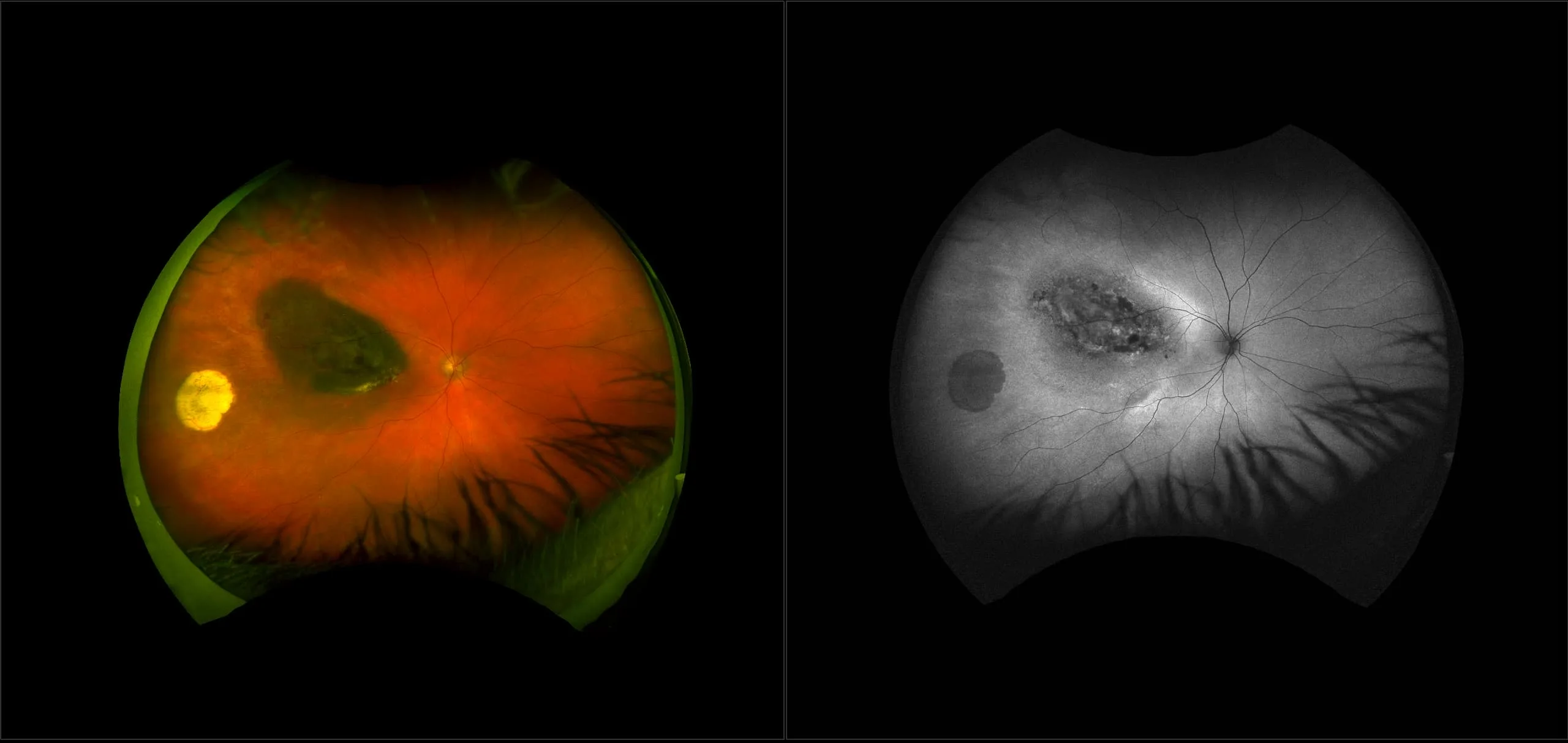
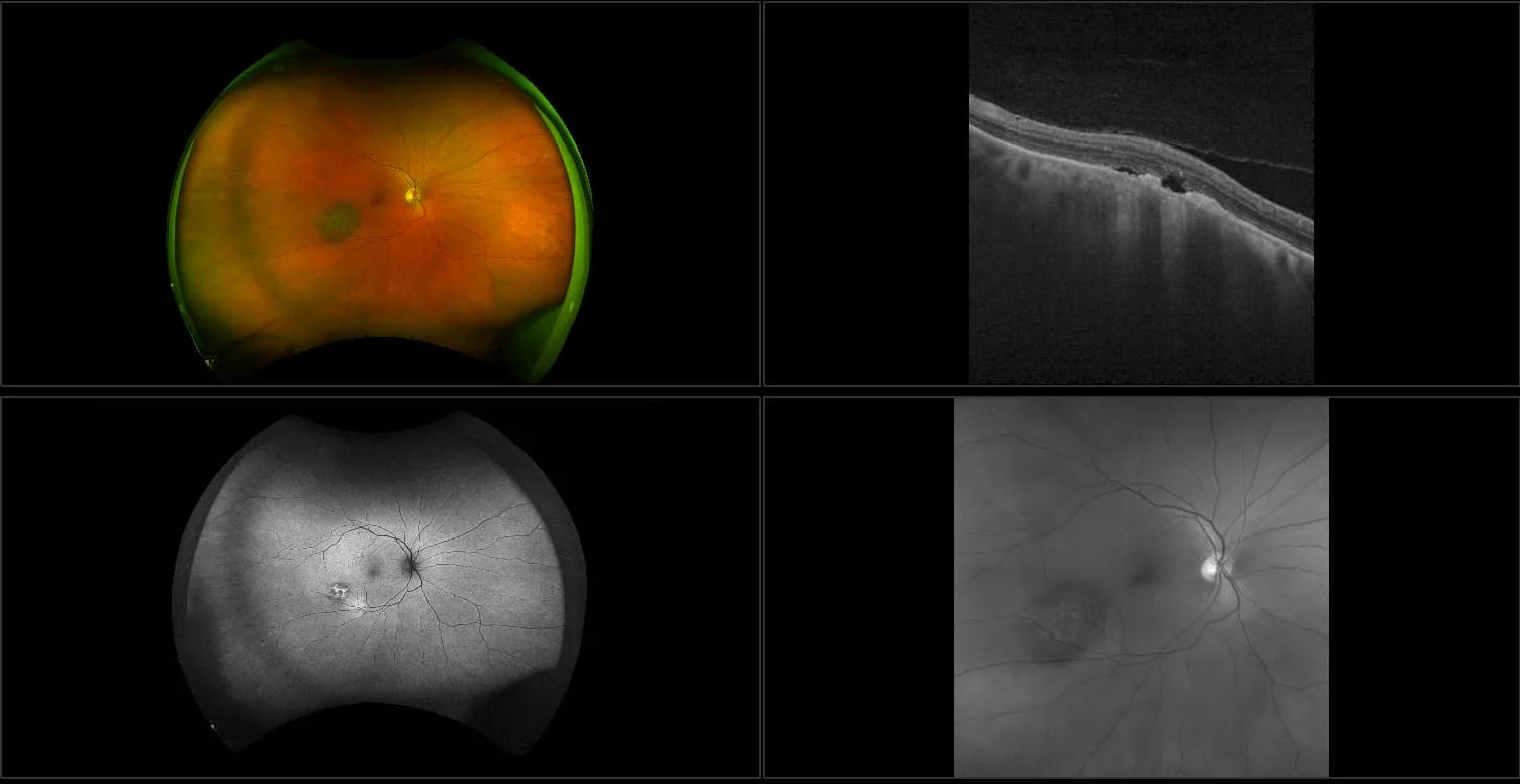
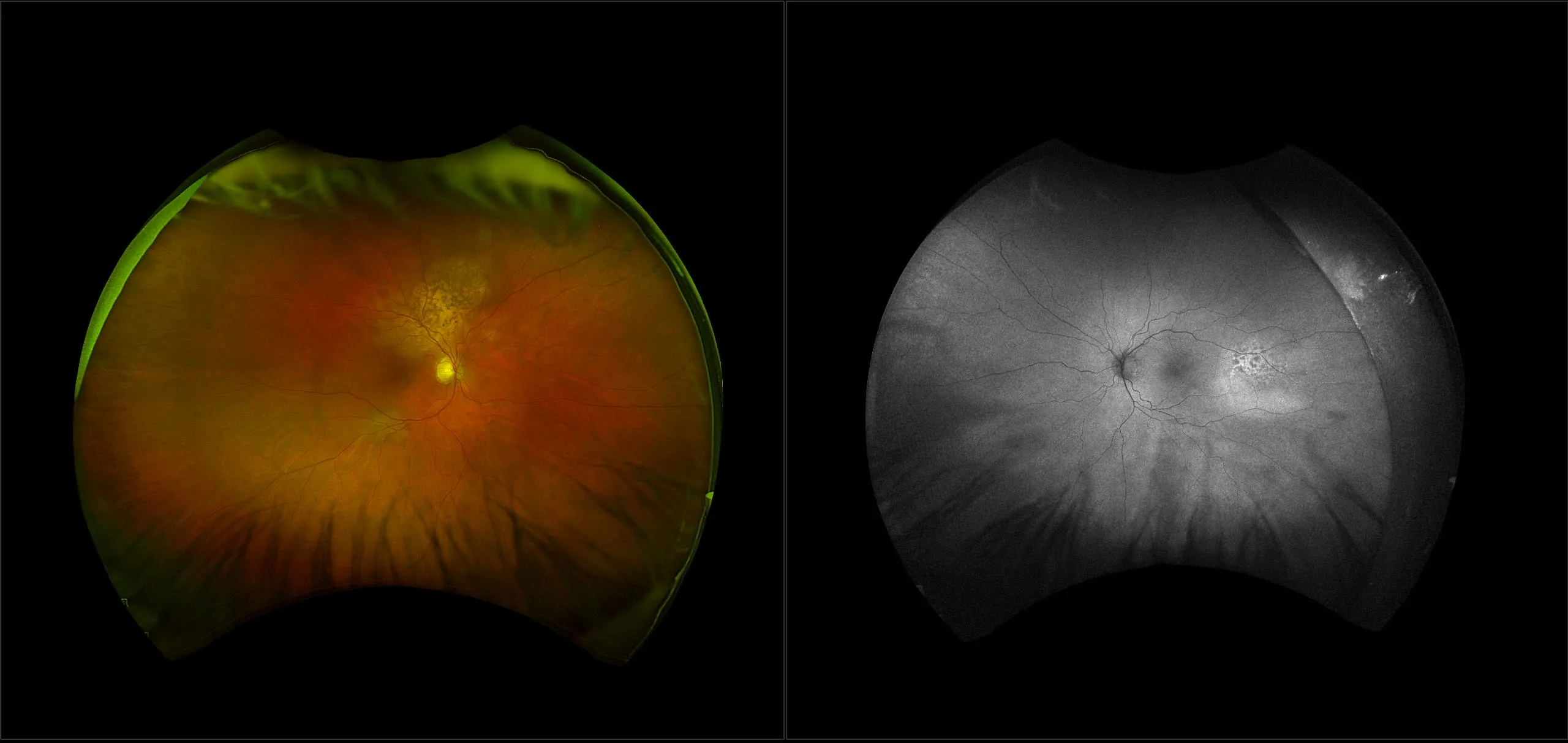
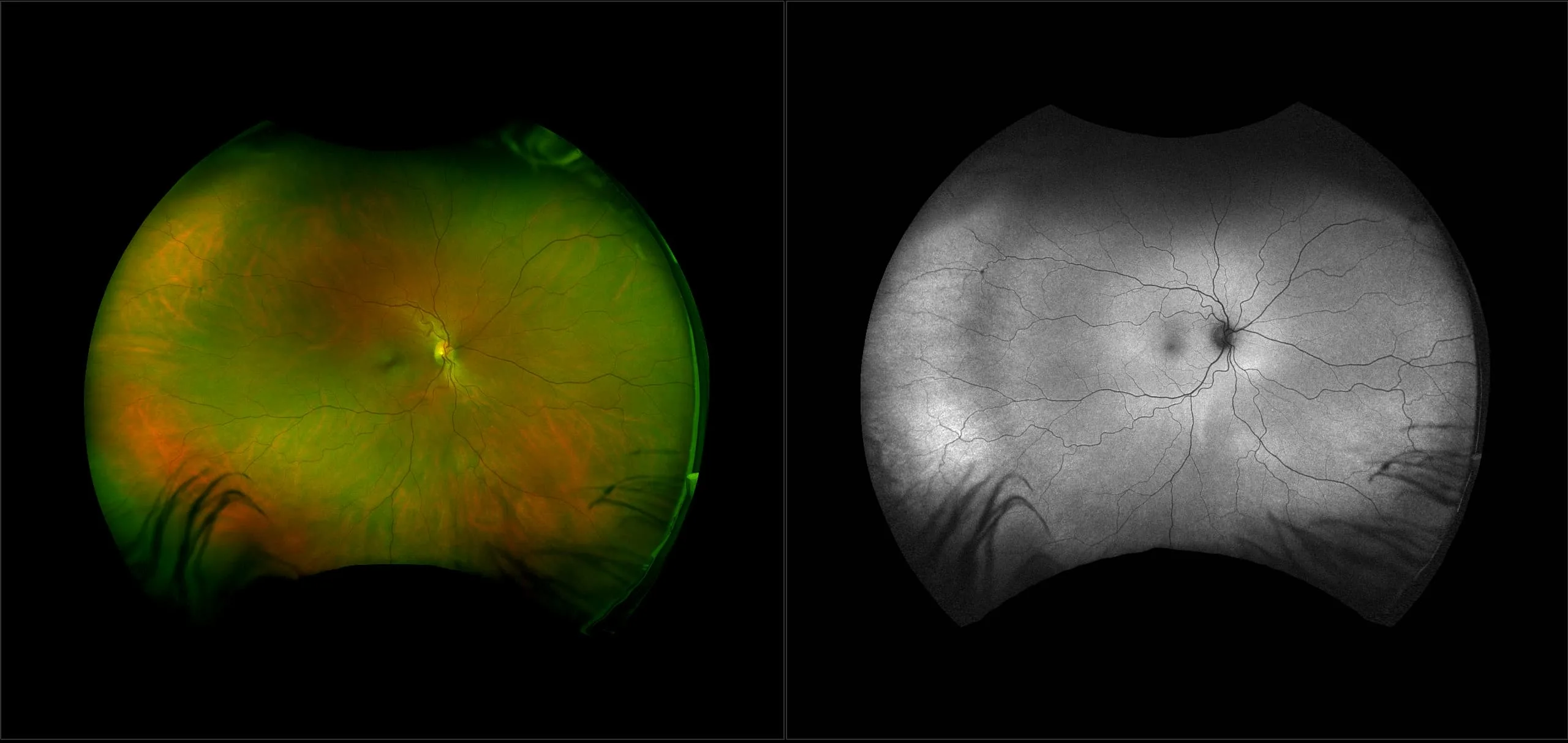
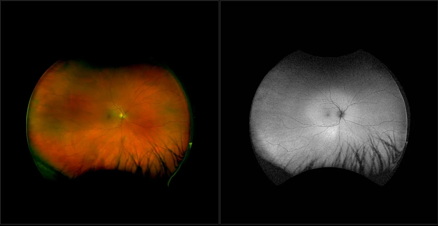
optomap multimodal imaging is transforming how eye care professionals approach ocular oncology by providing a high-resolution wider view of the retina and choroid. With the ability to uncover important details about ocular tumors and related retinal changes, optomap helps clinicians identify retinal lesions, plan more effective treatments, and follow patients over time with confidence. Fast, non-invasive, and cost-effective, optomap brings advanced technology into everyday practice, improving outcomes for patients facing complex ocular conditions.
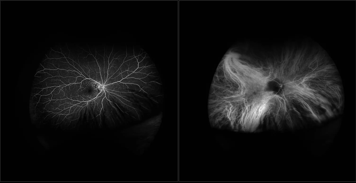
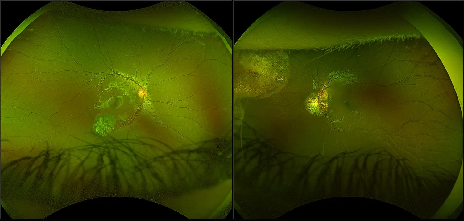
“With [ultra] widefield FA, smaller peripheral lesions that may not be visible on routine exams or fundus photography may be found sooner, allowing for earlier, less-invasive treatment with laser photocoagulation, which has minimal side effects and may halt progression of the lesions before vision loss occurs.”
Clinical Summary
optomap is More Informative and Cost Effective for Ocular Oncology
optomap provides more information than traditional CFP for diagnosis & management of ocular oncology.
Referenced Papers
Comparison of a Novel Ultra-Widefield Three-Color Scanning Laser Ophthalmoscope to Other Retinal Imaging Modalities in Chorioretinal Lesion Imaging.
Comparative Study of Clinical, Ultrasonographic, Conventional Imaging, and Ultra-Wide-Field Fundus for Measurements of the Longest Basal Diameter of Choroidal Tumors
Comparison of basal diameters in choroidal melanoma using ultra-widefield color fundus photography and ultrasonography
Pixel Intensity to Estimate Choroidal Tumor Thickness Using 2-Dimensional Ultra-Widefield Images.
Evaluation of OPTOS wide-field fundus image projections for radiotherapy planning of uveal melanoma
3D WrapTM Ultra-Widefield Reconstruction in Stereotactic Radiosurgery for Choroidal Melanoma
Early Detection of Retinal Hemangioblastomas in Von Hippel-Lindau Disease using Ultra-widefield Fluorescein Angiography.
Efficacy of Retinal Lesion Screening in Von Hippel-Lindau Patients With Widefield Color Fundus Imaging Versus Widefield FA
Intravascular large B-cell lymphoma of the eye: Literature review and new findings
Comparing the use of the Optos Silverstone widefield imaging system with ultrasound B-scanning for the assessment of choroidal naevi
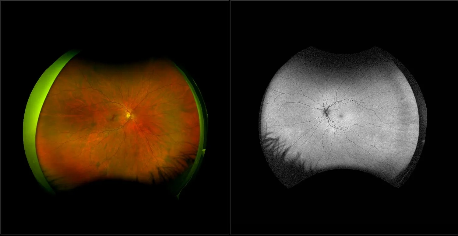
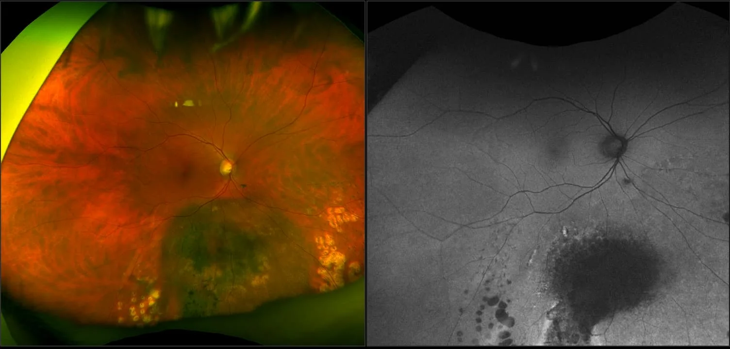
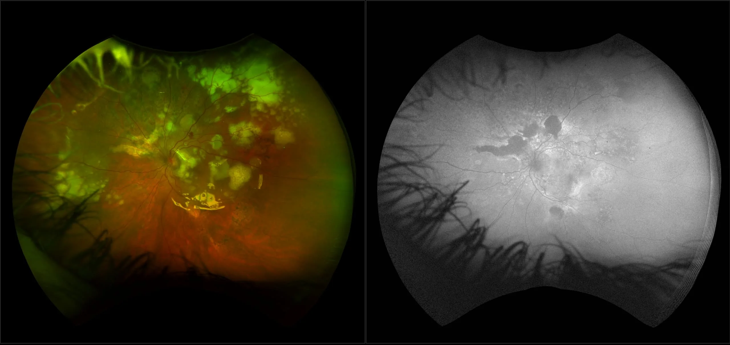
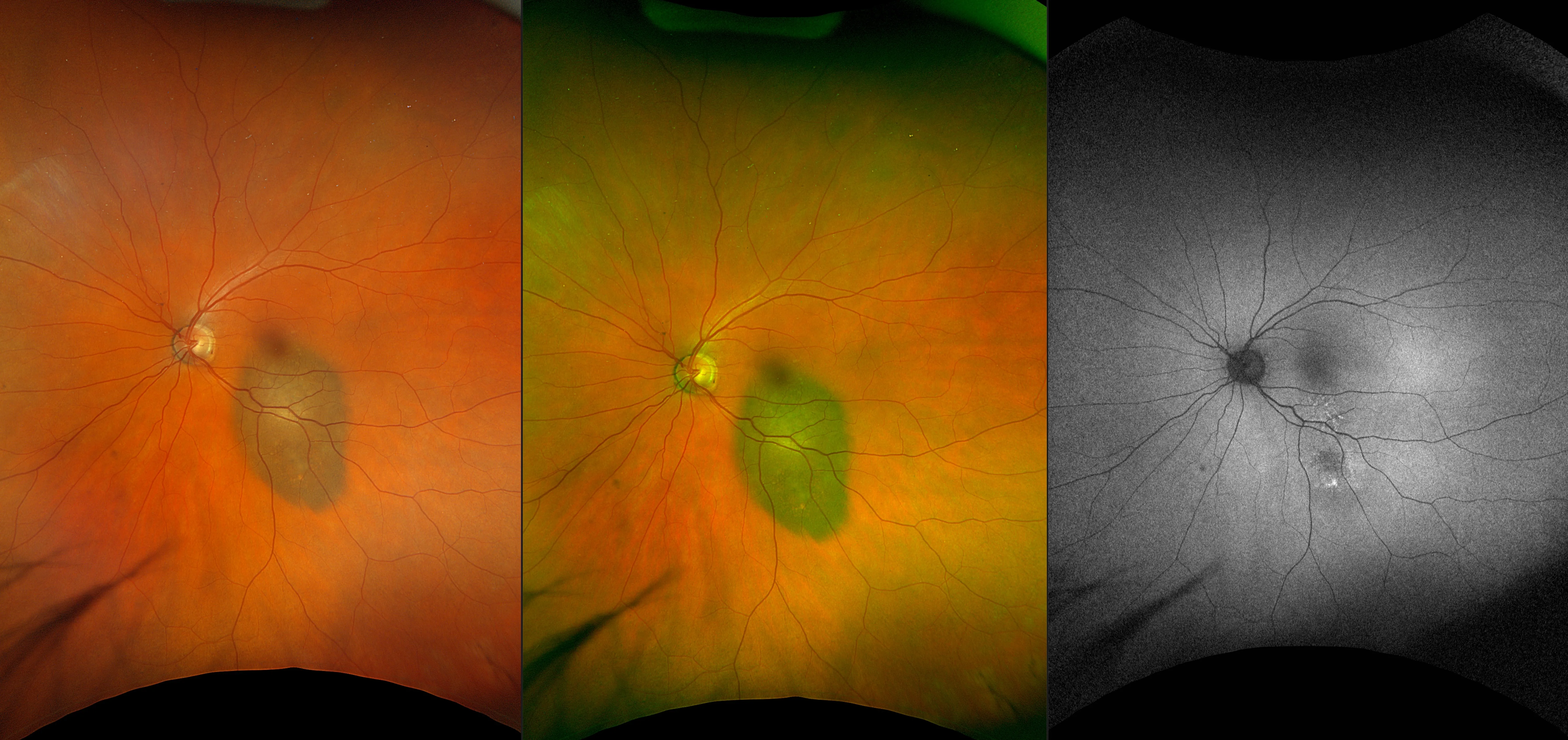
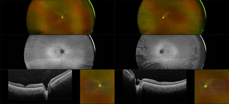
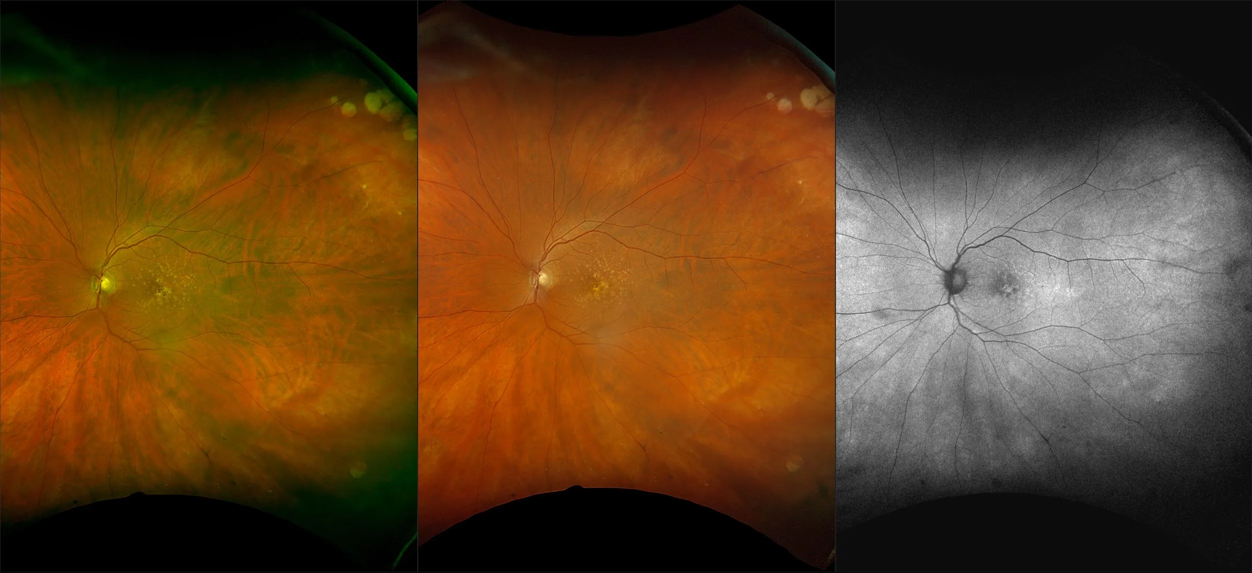
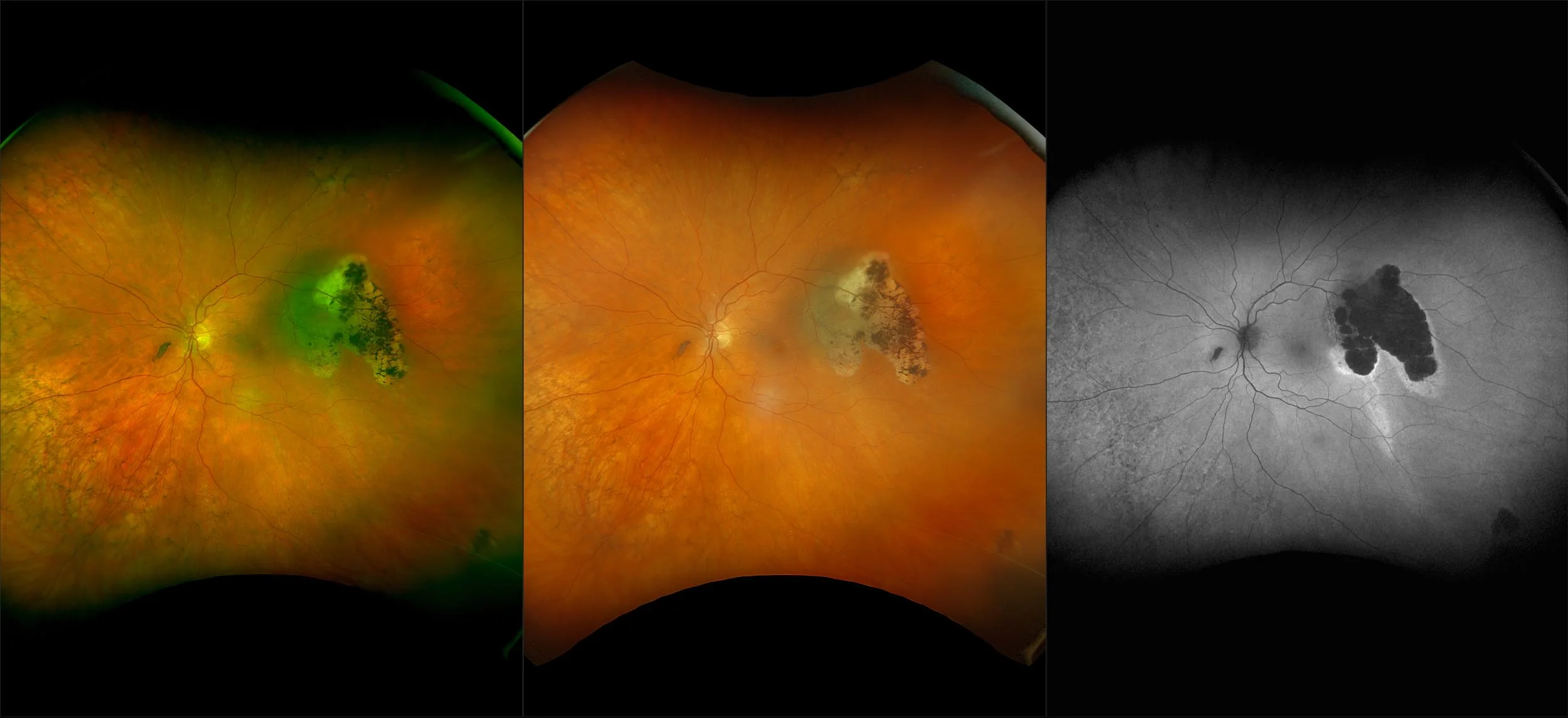
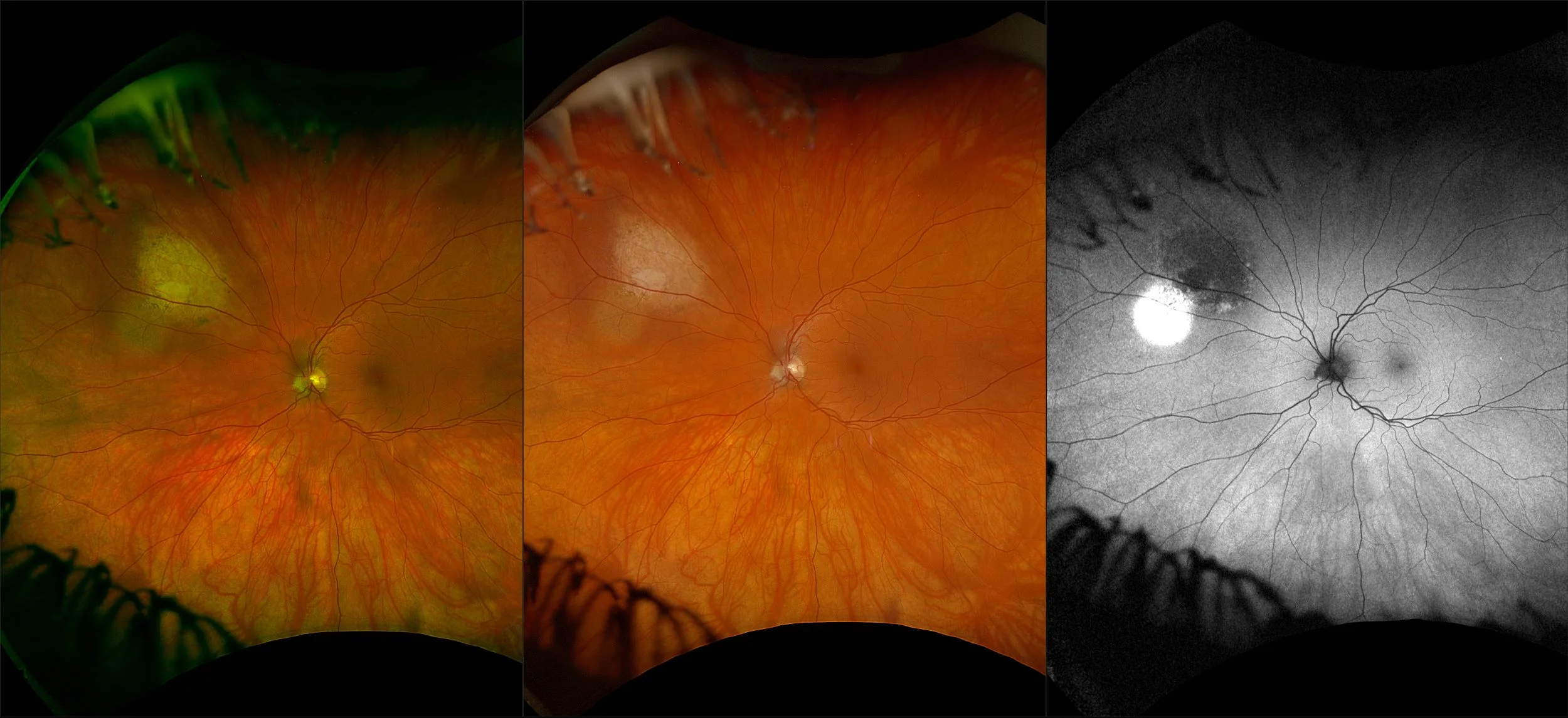
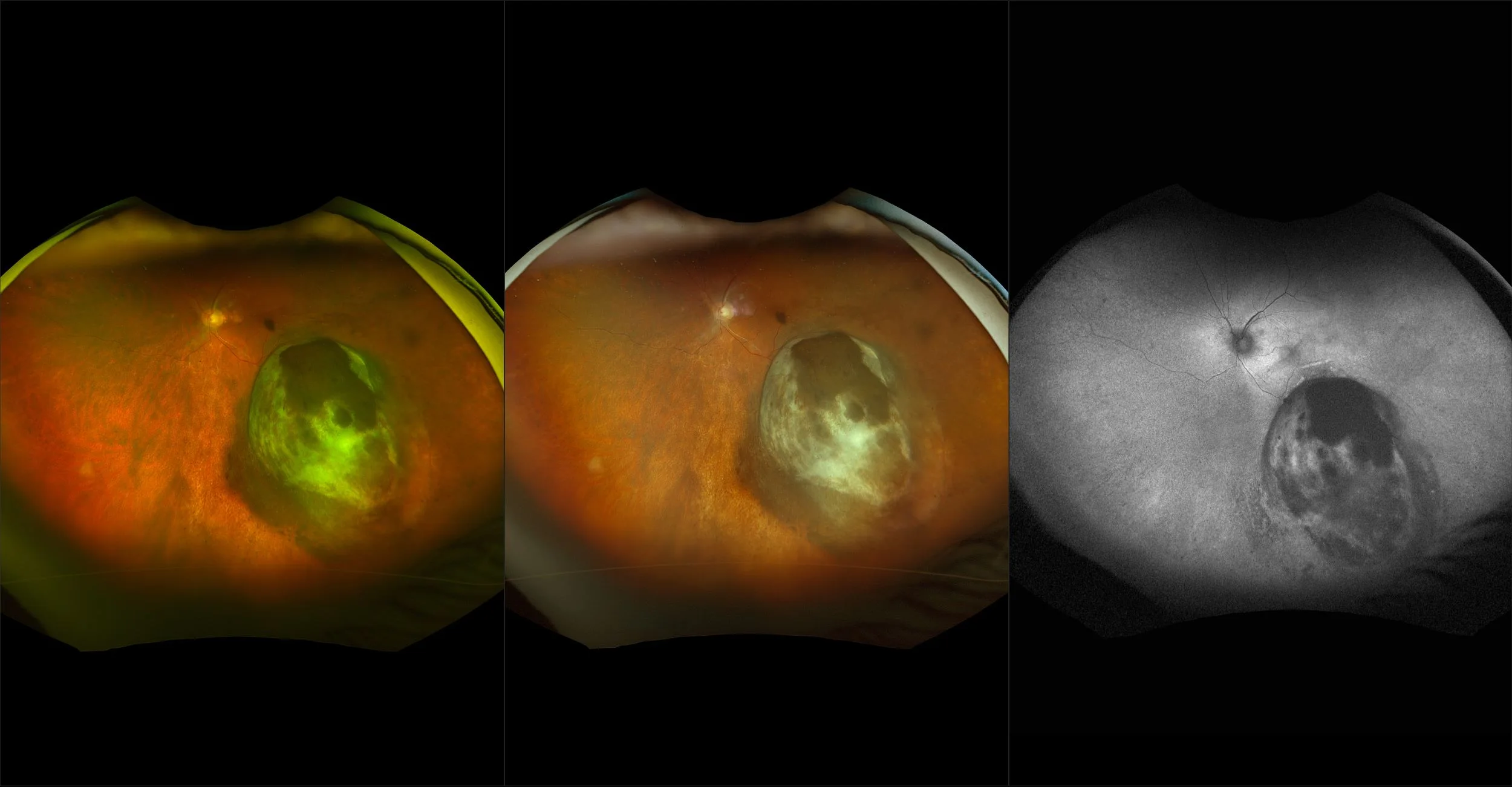
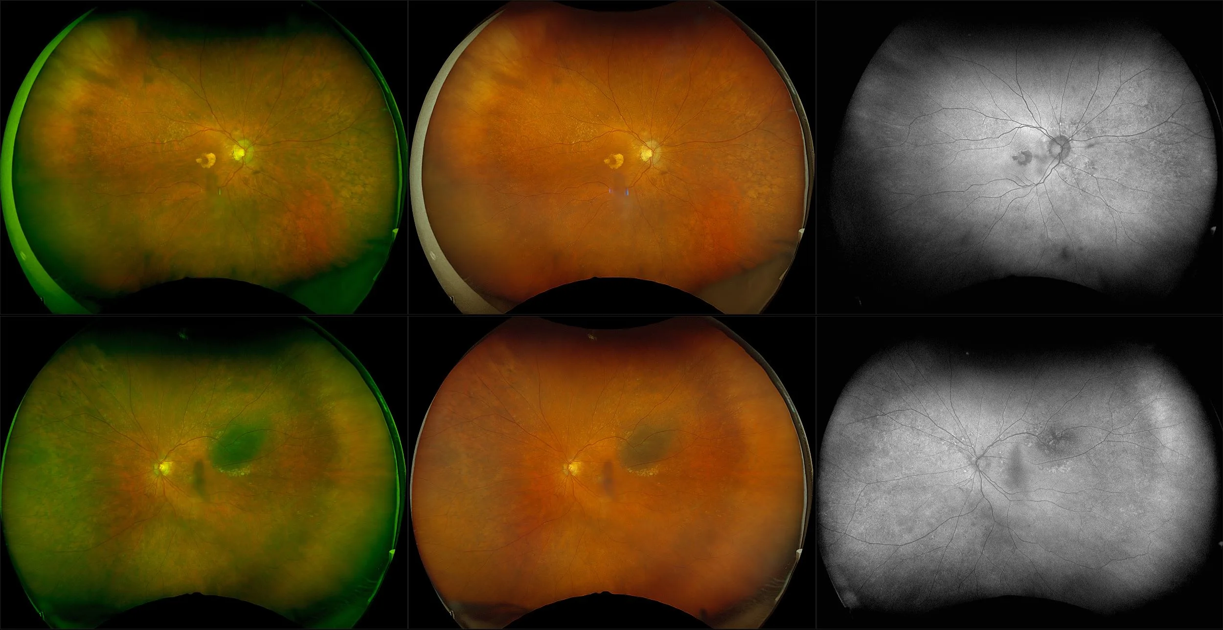
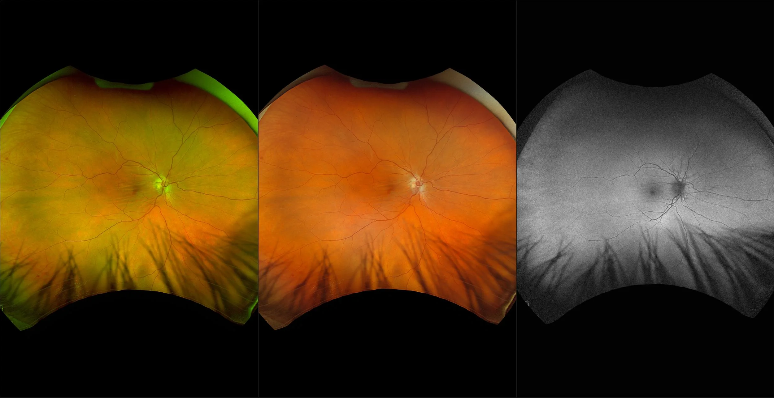
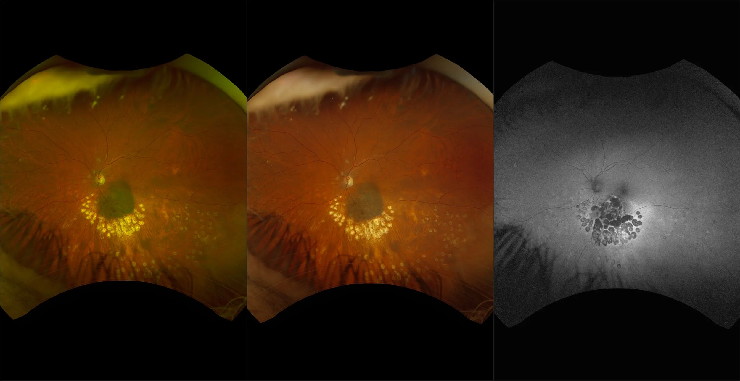
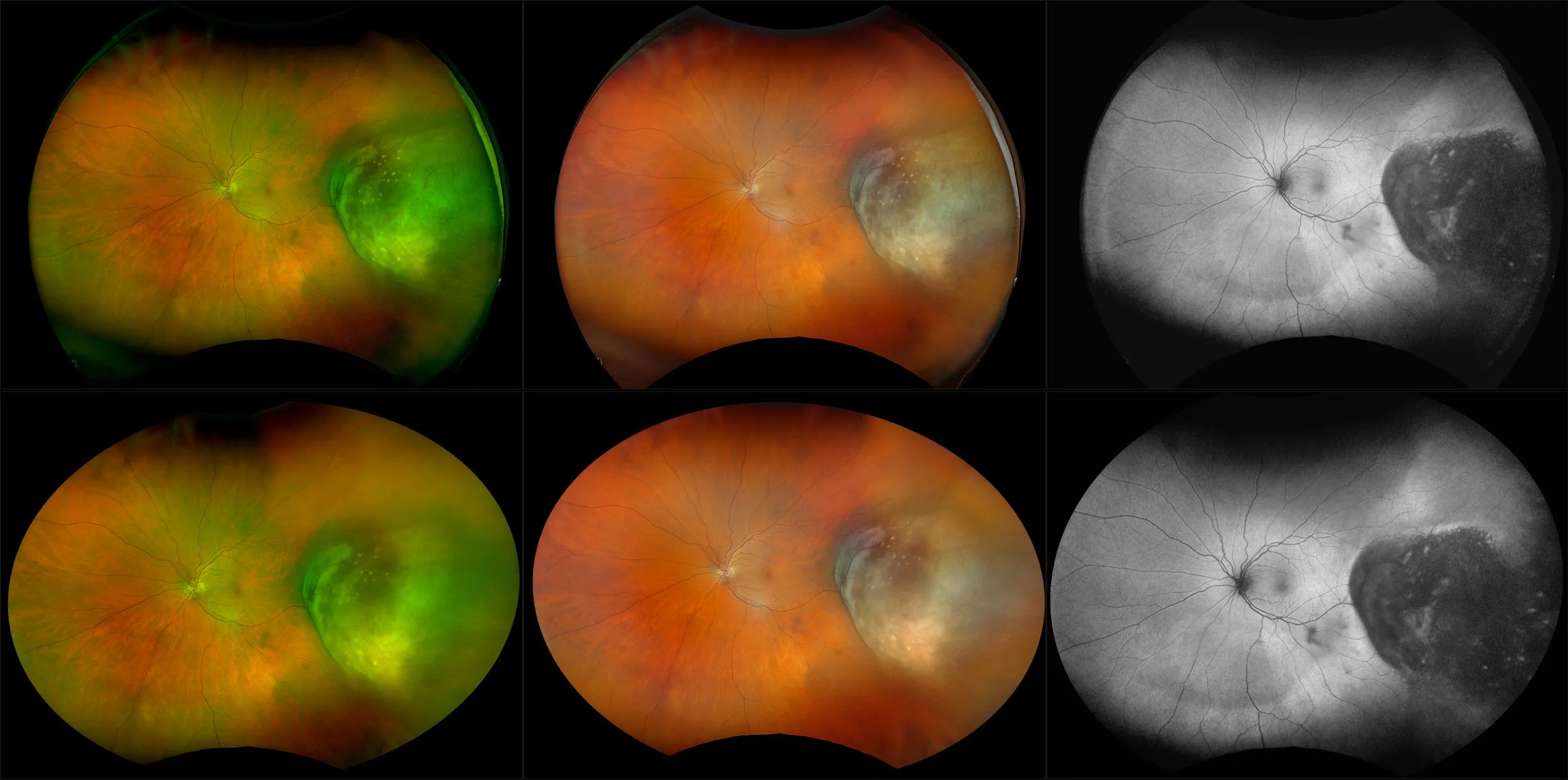
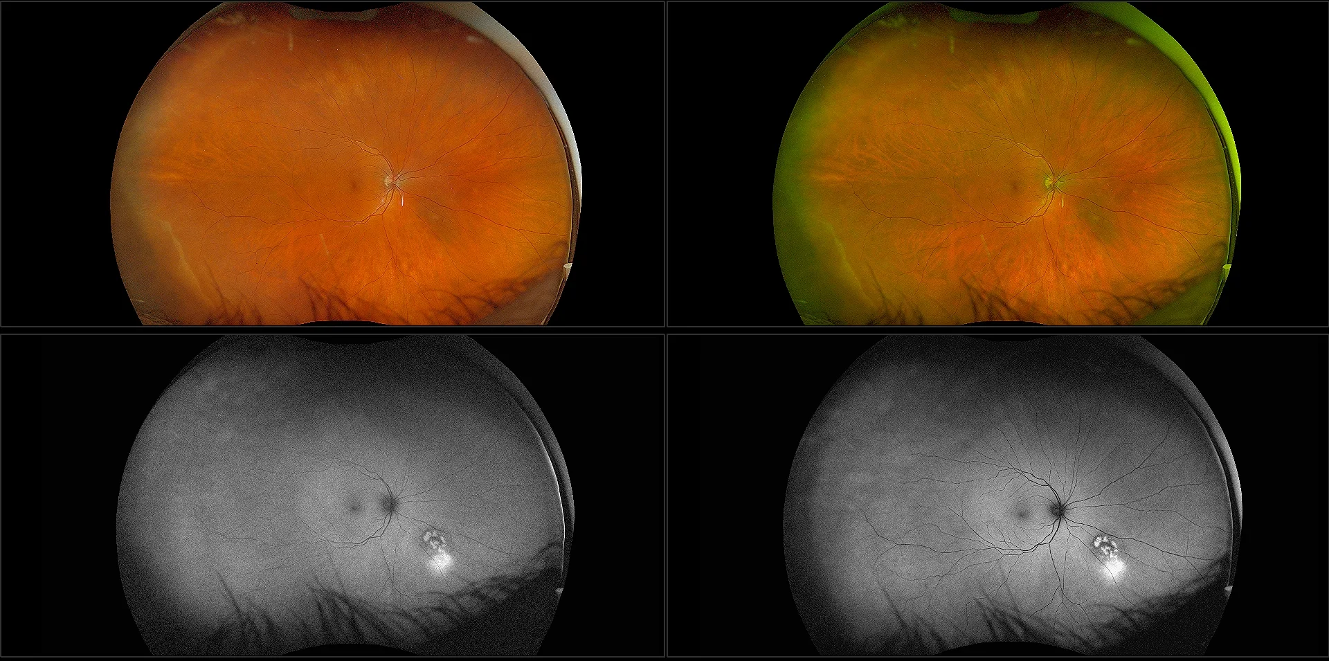
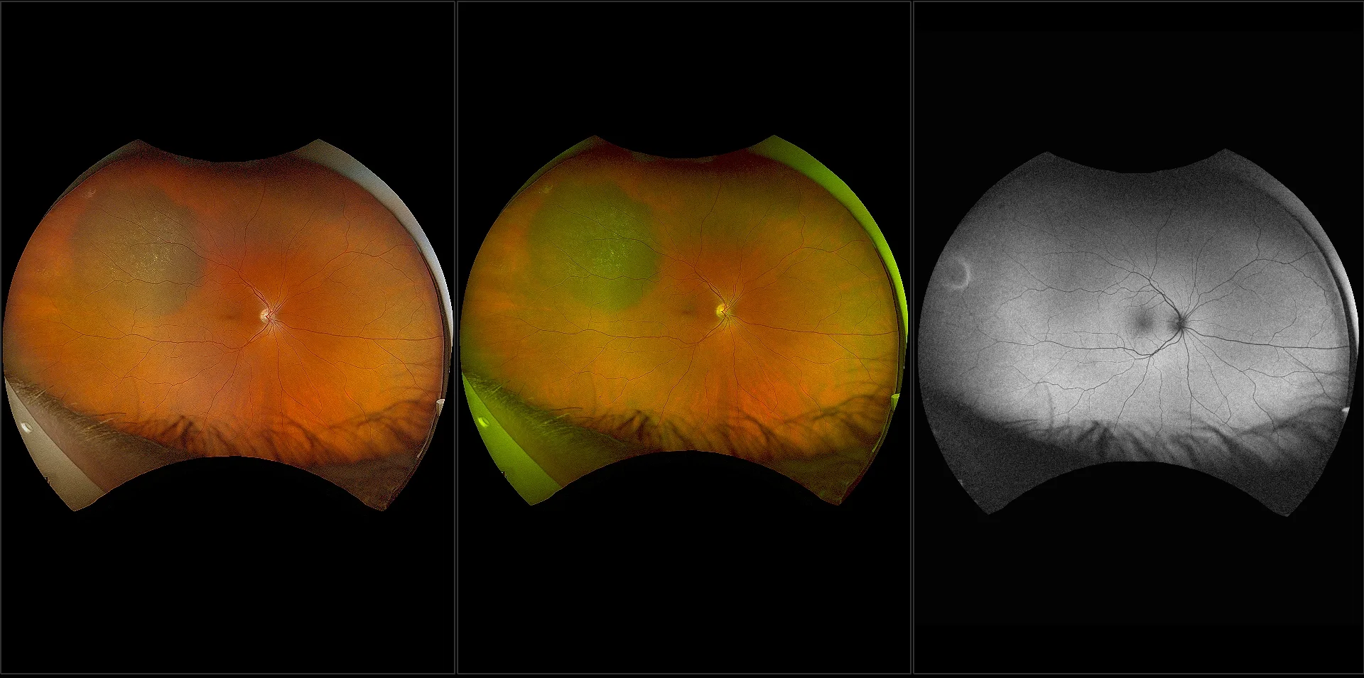
optomap Multimodal Ocular Oncology Cases
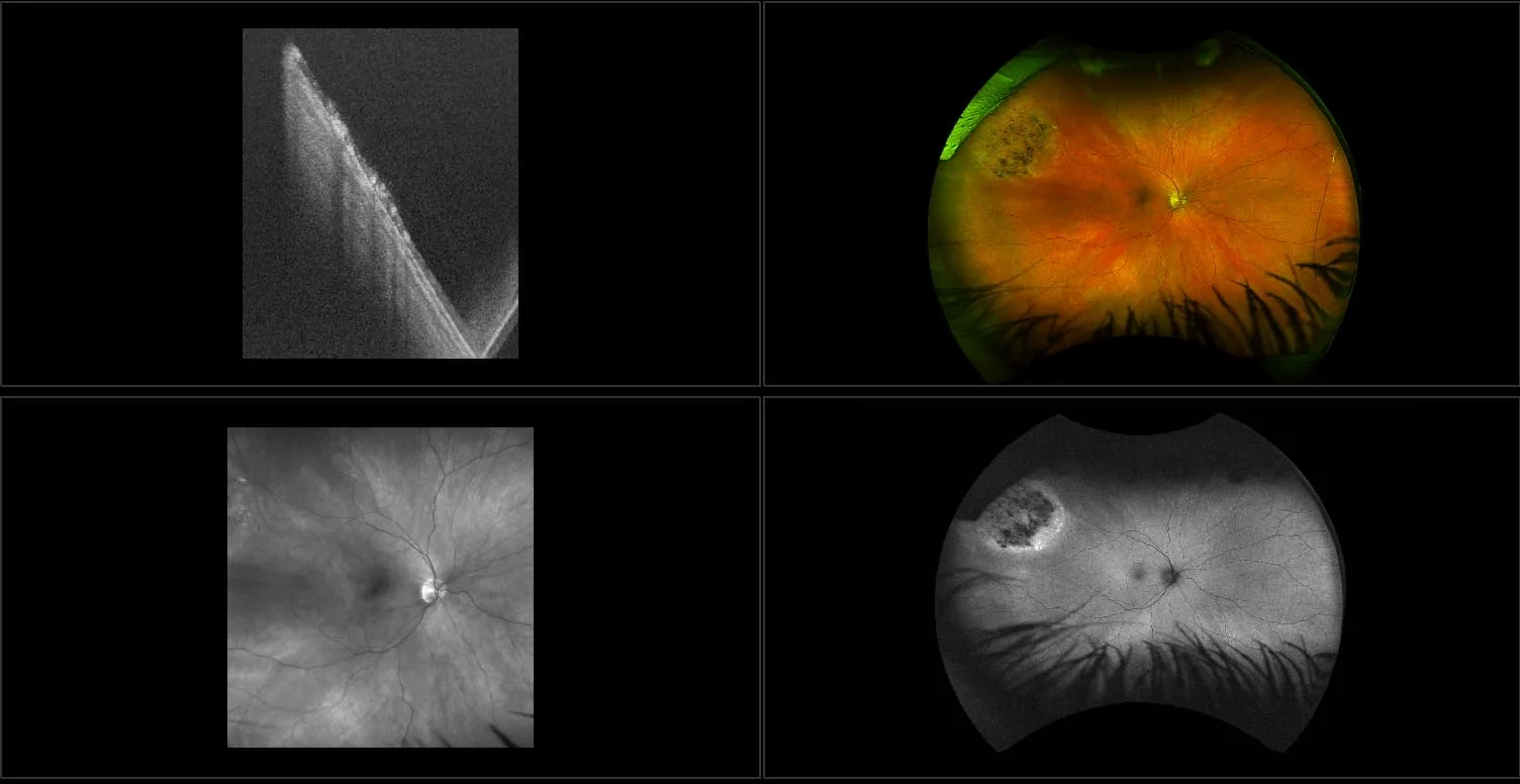
Optos offers multimodal imaging with all ultra-widefield devices. Having both ultra-widefield and four images captured in less than one second has been shown to enhance pathology detection and disease management as well as improve practice and clinic flow. Ultra-widefield multimodal imaging is important across all access points of patient care - screening, detection, diagnosis, and treatment.
Related Cases
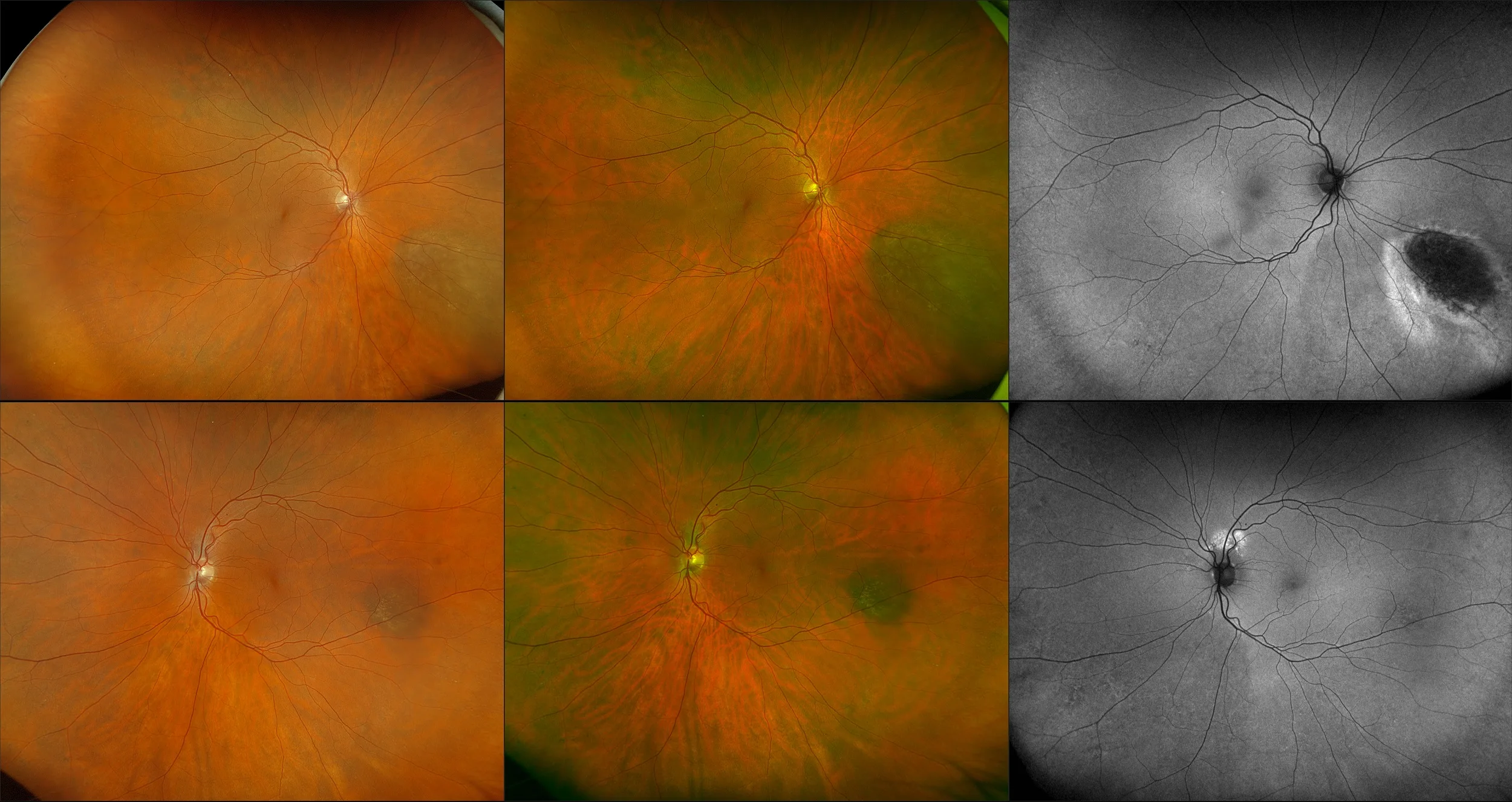

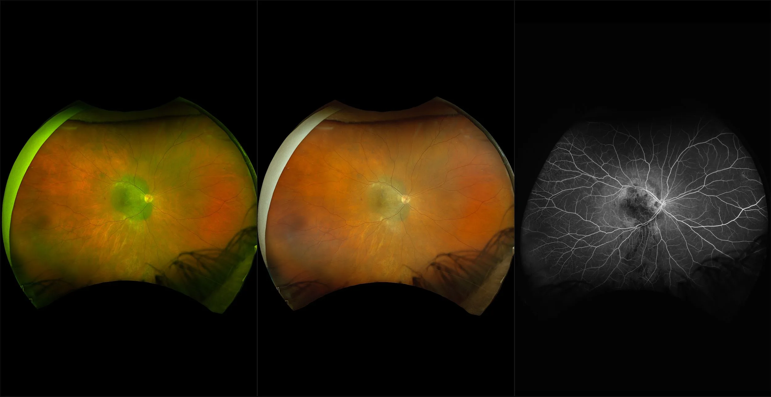
Ocular Oncology Care with optomap
Optos offers multimodal imaging with all ultra-widefield devices. Having both ultra-widefield and four images captured in less than one second has been shown to enhance pathology detection and disease management as well as improve practice and clinic flow. Ultra-widefield multimodal imaging is important across all access points of patient care - screening, detection, diagnosis, and treatment.


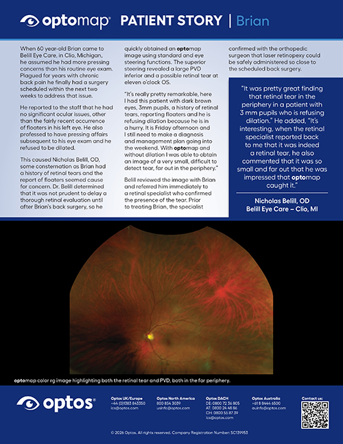
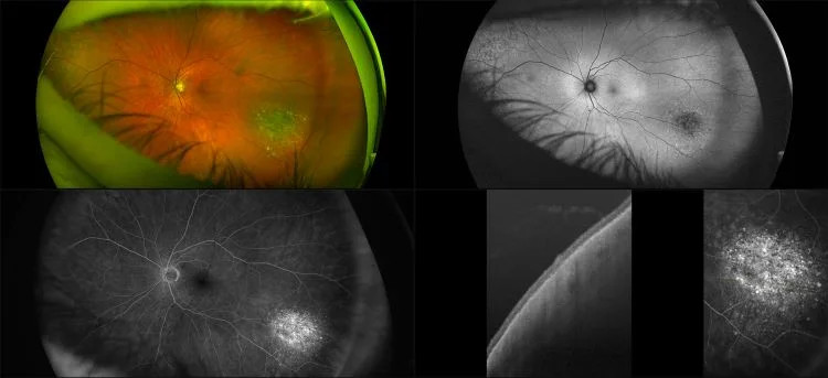
Patient Story - Brian
When 60 year-old Brian came to Belill Eye Care, in Clio, Michigan, for a non-symptomatic, routine eye exam, he never thought that his optomap would reveal that he needed to be sent for potentially sight-saving laser surgery for a tear far out in his periphery.